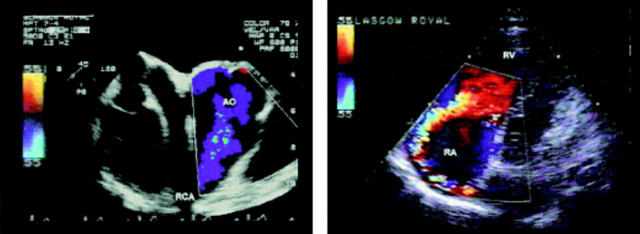
Figure 13 .
Images from a man with a fistula from the right coronary artery to the right atrium. The transoesophageal image (left) shows a very dilated right coronary artery which passes anteriorly and then sharply posteriorly. The transthoracic image (right) shows the jet flowing into the right atrium towards the right wall and then anteriorly; others as before.

