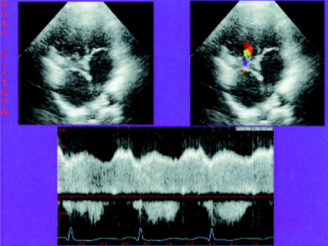
Figure 28 .
Transthoracic apical four chamber study of total cavopulmonary connection. The left image shows a double inlet ventricle and the proximity of the intra-atrial conduit to the right valve and the right shows colour flow through a surgically created fenestration and then through the atrioventricular valve to the ventricular chamber. The spectral Doppler trace of flow through the fenestration (below) shows right to left flow which is continuous and of relatively high velocity.

