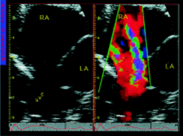
Figure 3 .
Cross sectional and colour images of a high sinus venosus atrial septal defect. The scanning plane has been tilted upwards to show the entry of the superior vena cava, which is shown to override the atrial septum. An anomalous pulmonary vein is not shown. SVC, superior vena cava; others as before.

