Full Text
The Full Text of this article is available as a PDF (344.1 KB).
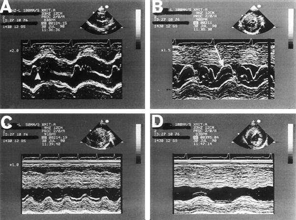
Figure 1 .
Typical M mode echocardiogram from a patient with hypertrophic cardiomyopathy highlighting the four main echocardiographic features of the condition. (A) Midsystolic closure of the aortic valve (arrowhead); (B) systolic anterior motion of the mitral valve (arrow) and asymmetric left ventricular hypertrophy together with a small, vigorously contracting left ventricle. In frames (C) and (D), the M mode beam passes through the septum and posterior wall beyond the mitral valve, at the level of the papillary muscles and apex, demonstrating the large reduction of left ventricular end systolic dimensions. Reproduced from Nihoyannopoulos and McKenna with permission of Churchill Livingstone.10
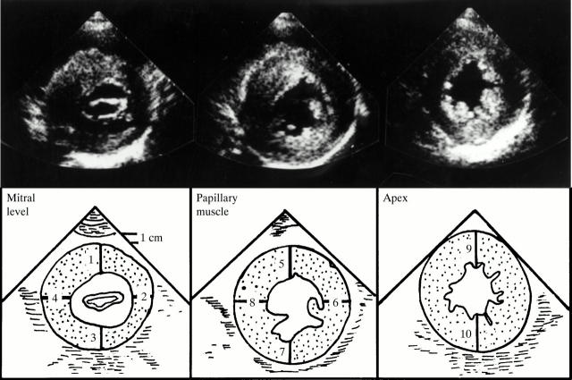
Figure 2 .
An example of serial short axis, cross sectional views of the left ventricle at three levels—the mitral valve, papillary muscles, and apex—demonstrating the segments of myocardial wall measured routinely in patients with hypertrophic cardiomyopathy in our laboratory. Reproduced from Nihoyannopoulos and McKenna with permission of Churchill Livingstone.10
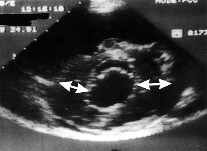
Figure 3 .
Parasternal short axis view of the left ventricle at the mitral valve level demonstrating a typical eccentric form of ventricular hypertrophy localised essentially at the lateral wall and posterior septum, while the anterior and posterior walls are normal. Reproduced from Nihoyannopoulos and McKenna with permission of Churchill Livingstone.10
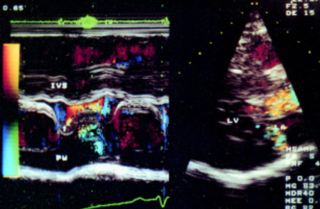
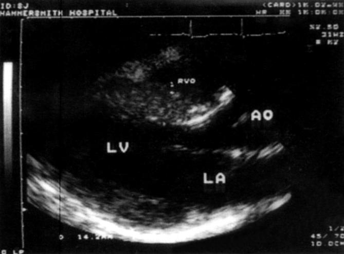
Figure 4 .
Parasternal long axis view with colour M mode Doppler echocardiography from a patient with hypertrophic cardiomyopathy and a high (90 mmHg) outflow tract gradient. Notice the occurrence of systolic anterior motion of the mitral valve at the same time as the presence of outflow turbulence (arrow), and later into systole, the presence of mitral regurgitation.
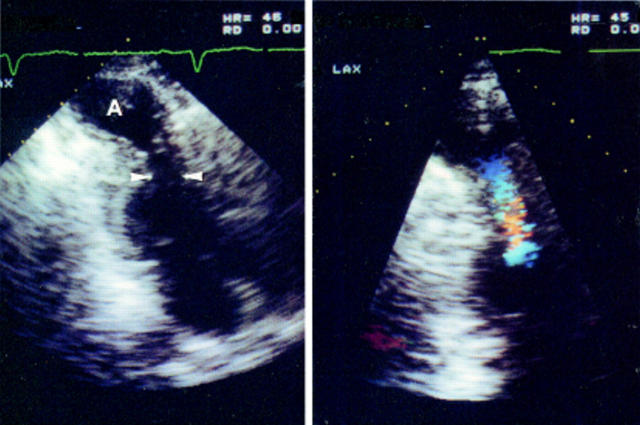
Figure 5 .
Apical long axis view with colour flow mapping in a patient with hypertrophic cardiomyopathy and a midventricular gradient and an apical aneurysm. Note, on the left, a midventricular narrowing of the left ventricular cavity (arrowheads) and, on the right, the turbulent flow on colour Doppler echocardiography originating at that level. A, apical aneurysm.
Figure 6 .
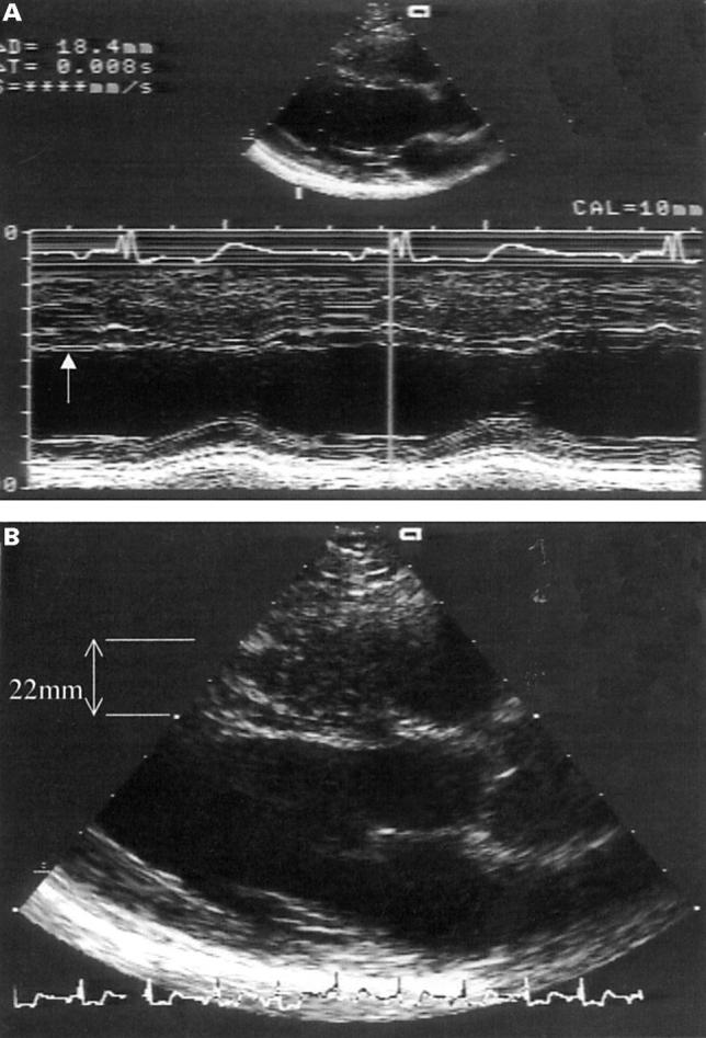
Parasternal long axis view from a patient referred with the echocardiographic diagnosis of hypertrophic cardiomyopathy (HCM). (A) The M mode image showing the normal septal thickness (10 mm). Notice that the measurement excludes the false tendon (arrow), which runs parallel to the septum. (B) The false tendon along the ventricular septum, which was included in the original measurement of 22 mm and led to the wrong diagnosis of HCM.
Figure 7 .
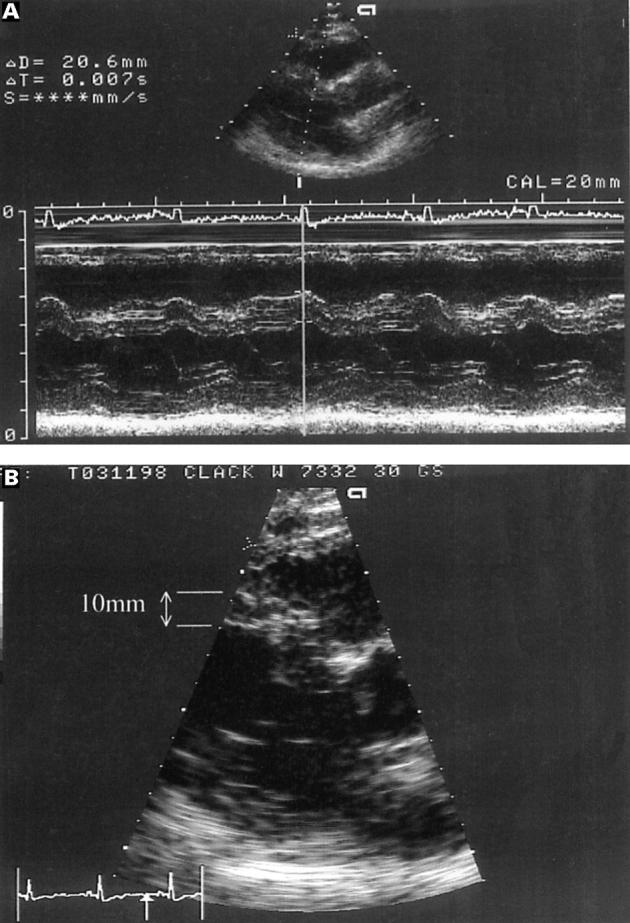
Parasternal long axis view from a patient referred with the echocardiographic diagnosis of hypertrophic cardiomyopathy. (A) The M mode image showing the original measurement of 20 mm for the ventricular septum passing obliquely through the angulated (sigmoid) septum. (B) The two dimensional picture with the angulated septum showing the correct measurement of the septum (10 mm).
Figure 8 .
Parasternal long axis from a patient with Friedreich's ataxia demonstrating asymmetric septal hypertrophy. Reproduced with permission from Dutka et al.29
Selected References
These references are in PubMed. This may not be the complete list of references from this article.
- Alfonso F., Nihoyannopoulos P., Stewart J., Dickie S., Lemery R., McKenna W. J. Clinical significance of giant negative T waves in hypertrophic cardiomyopathy. J Am Coll Cardiol. 1990 Apr;15(5):965–971. doi: 10.1016/0735-1097(90)90225-e. [DOI] [PubMed] [Google Scholar]
- Appleton C. P., Hatle L. K., Popp R. L. Relation of transmitral flow velocity patterns to left ventricular diastolic function: new insights from a combined hemodynamic and Doppler echocardiographic study. J Am Coll Cardiol. 1988 Aug;12(2):426–440. doi: 10.1016/0735-1097(88)90416-0. [DOI] [PubMed] [Google Scholar]
- Bonne G., Carrier L., Bercovici J., Cruaud C., Richard P., Hainque B., Gautel M., Labeit S., James M., Beckmann J. Cardiac myosin binding protein-C gene splice acceptor site mutation is associated with familial hypertrophic cardiomyopathy. Nat Genet. 1995 Dec;11(4):438–440. doi: 10.1038/ng1295-438. [DOI] [PubMed] [Google Scholar]
- Brenner J. I., Baker K., Ringel R. E., Berman M. A. Echocardiographic evidence of left ventricular bands in infants and children. J Am Coll Cardiol. 1984 Jun;3(6):1515–1520. doi: 10.1016/s0735-1097(84)80291-0. [DOI] [PubMed] [Google Scholar]
- Charron P., Dubourg O., Desnos M., Isnard R., Hagege A., Millaire A., Carrier L., Bonne G., Tesson F., Richard P. Diagnostic value of electrocardiography and echocardiography for familial hypertrophic cardiomyopathy in a genotyped adult population. Circulation. 1997 Jul 1;96(1):214–219. doi: 10.1161/01.cir.96.1.214. [DOI] [PubMed] [Google Scholar]
- Doi Y. L., McKenna W. J., Gehrke J., Oakley C. M., Goodwin J. F. M mode echocardiography in hypertrophic cardiomyopathy: diagnostic criteria and prediction of obstruction. Am J Cardiol. 1980 Jan;45(1):6–14. doi: 10.1016/0002-9149(80)90213-1. [DOI] [PubMed] [Google Scholar]
- Dutka D. P., Donnelly J. E., Nihoyannopoulos P., Oakley C. M., Nunez D. J. Marked variation in the cardiomyopathy associated with Friedreich's ataxia. Heart. 1999 Feb;81(2):141–147. doi: 10.1136/hrt.81.2.141. [DOI] [PMC free article] [PubMed] [Google Scholar]
- Goor D., Lillehei C. W., Edwards J. E. The "sigmoid septum". Variation in the contour of the left ventricular outt. Am J Roentgenol Radium Ther Nucl Med. 1969 Oct;107(2):366–376. [PubMed] [Google Scholar]
- Goor D., Lillehei C. W., Edwards J. E. The "sigmoid septum". Variation in the contour of the left ventricular outt. Am J Roentgenol Radium Ther Nucl Med. 1969 Oct;107(2):366–376. [PubMed] [Google Scholar]
- Hanrath P., Mathey D. G., Siegert R., Bleifeld W. Left ventricular relaxation and filling pattern in different forms of left ventricular hypertrophy: an echocardiographic study. Am J Cardiol. 1980 Jan;45(1):15–23. doi: 10.1016/0002-9149(80)90214-3. [DOI] [PubMed] [Google Scholar]
- Klues H. G., Schiffers A., Maron B. J. Phenotypic spectrum and patterns of left ventricular hypertrophy in hypertrophic cardiomyopathy: morphologic observations and significance as assessed by two-dimensional echocardiography in 600 patients. J Am Coll Cardiol. 1995 Dec;26(7):1699–1708. doi: 10.1016/0735-1097(95)00390-8. [DOI] [PubMed] [Google Scholar]
- Krasnow N. Subaortic septal bulge simulates hypertrophic cardiomyopathy by angulation of the septum with age, independent of focal hypertrophy. An echocardiographic study. J Am Soc Echocardiogr. 1997 Jun;10(5):545–555. doi: 10.1016/s0894-7317(97)70009-9. [DOI] [PubMed] [Google Scholar]
- Lewis J. F., Maron B. J. Diversity of patterns of hypertrophy in patients with systemic hypertension and marked left ventricular wall thickening. Am J Cardiol. 1990 Apr 1;65(13):874–881. doi: 10.1016/0002-9149(90)91429-a. [DOI] [PubMed] [Google Scholar]
- Maron B. J., Bonow R. O., Cannon R. O., 3rd, Leon M. B., Epstein S. E. Hypertrophic cardiomyopathy. Interrelations of clinical manifestations, pathophysiology, and therapy (1). N Engl J Med. 1987 Mar 26;316(13):780–789. doi: 10.1056/NEJM198703263161305. [DOI] [PubMed] [Google Scholar]
- Maron B. J., Clark C. E., Henry W. L., Fukuda T., Edwards J. E., Mathews E. C., Jr, Redwood D. R., Epstein S. E. Prevalence and characteristics of disproportionate ventricular septal thickening in patients with acquired or congenital heart diseases: echocardiographic and morphologic findings. Circulation. 1977 Mar;55(3):489–496. doi: 10.1161/01.cir.55.3.489. [DOI] [PubMed] [Google Scholar]
- Maron B. J., Gottdiener J. S., Epstein S. E. Patterns and significance of distribution of left ventricular hypertrophy in hypertrophic cardiomyopathy. A wide angle, two dimensional echocardiographic study of 125 patients. Am J Cardiol. 1981 Sep;48(3):418–428. doi: 10.1016/0002-9149(81)90068-0. [DOI] [PubMed] [Google Scholar]
- Maron B. J., Nichols P. F., 3rd, Pickle L. W., Wesley Y. E., Mulvihill J. J. Patterns of inheritance in hypertrophic cardiomyopathy: assessment by M-mode and two-dimensional echocardiography. Am J Cardiol. 1984 Apr 1;53(8):1087–1094. doi: 10.1016/0002-9149(84)90643-x. [DOI] [PubMed] [Google Scholar]
- Maron B. J., Spirito P., Green K. J., Wesley Y. E., Bonow R. O., Arce J. Noninvasive assessment of left ventricular diastolic function by pulsed Doppler echocardiography in patients with hypertrophic cardiomyopathy. J Am Coll Cardiol. 1987 Oct;10(4):733–742. doi: 10.1016/s0735-1097(87)80264-4. [DOI] [PubMed] [Google Scholar]
- McKenna W. J., Spirito P., Desnos M., Dubourg O., Komajda M. Experience from clinical genetics in hypertrophic cardiomyopathy: proposal for new diagnostic criteria in adult members of affected families. Heart. 1997 Feb;77(2):130–132. doi: 10.1136/hrt.77.2.130. [DOI] [PMC free article] [PubMed] [Google Scholar]
- McKenna W. J., Stewart J. T., Nihoyannopoulos P., McGinty F., Davies M. J. Hypertrophic cardiomyopathy without hypertrophy: two families with myocardial disarray in the absence of increased myocardial mass. Br Heart J. 1990 May;63(5):287–290. doi: 10.1136/hrt.63.5.287. [DOI] [PMC free article] [PubMed] [Google Scholar]
- Moolman J. C., Corfield V. A., Posen B., Ngumbela K., Seidman C., Brink P. A., Watkins H. Sudden death due to troponin T mutations. J Am Coll Cardiol. 1997 Mar 1;29(3):549–555. doi: 10.1016/s0735-1097(96)00530-x. [DOI] [PubMed] [Google Scholar]
- Nihoyannopoulos P., Karatasakis G., Frenneaux M., McKenna W. J., Oakley C. M. Diastolic function in hypertrophic cardiomyopathy: relation to exercise capacity. J Am Coll Cardiol. 1992 Mar 1;19(3):536–540. doi: 10.1016/s0735-1097(10)80268-2. [DOI] [PubMed] [Google Scholar]
- Pierard L. A., Henrard L., Noel J. F. Detection of left ventricular false tendons by two-dimensional echocardiography. Acta Cardiol. 1985;40(2):229–235. [PubMed] [Google Scholar]
- Richardson P., McKenna W., Bristow M., Maisch B., Mautner B., O'Connell J., Olsen E., Thiene G., Goodwin J., Gyarfas I. Report of the 1995 World Health Organization/International Society and Federation of Cardiology Task Force on the Definition and Classification of cardiomyopathies. Circulation. 1996 Mar 1;93(5):841–842. doi: 10.1161/01.cir.93.5.841. [DOI] [PubMed] [Google Scholar]
- Sanderson J. E., Traill T. A., Sutton M. G., Brown D. J., Gibson D. G., Goodwin J. F. Left ventricular relaxation and filling in hypertrophic cardiomyopathy. An echocardiographic study. Br Heart J. 1978 Jun;40(6):596–601. doi: 10.1136/hrt.40.6.596. [DOI] [PMC free article] [PubMed] [Google Scholar]
- Shapiro L. M., McKenna W. J. Distribution of left ventricular hypertrophy in hypertrophic cardiomyopathy: a two-dimensional echocardiographic study. J Am Coll Cardiol. 1983 Sep;2(3):437–444. doi: 10.1016/s0735-1097(83)80269-1. [DOI] [PubMed] [Google Scholar]
- Spirito P., Pelliccia A., Proschan M. A., Granata M., Spataro A., Bellone P., Caselli G., Biffi A., Vecchio C., Maron B. J. Morphology of the "athlete's heart" assessed by echocardiography in 947 elite athletes representing 27 sports. Am J Cardiol. 1994 Oct 15;74(8):802–806. doi: 10.1016/0002-9149(94)90439-1. [DOI] [PubMed] [Google Scholar]
- Sutton M. G., Tajik A. J., Gibson D. G., Brown D. J., Seward J. B., Guiliani E. R. Echocardiographic assessment of left ventricular filling and septal and posterior wall dynamics in idiopathic hypertrophic subaortic stenosis. Circulation. 1978 Mar;57(3):512–520. doi: 10.1161/01.cir.57.3.512. [DOI] [PubMed] [Google Scholar]
- Takenaka K., Dabestani A., Gardin J. M., Russell D., Clark S., Allfie A., Henry W. L. Left ventricular filling in hypertrophic cardiomyopathy: a pulsed Doppler echocardiographic study. J Am Coll Cardiol. 1986 Jun;7(6):1263–1271. doi: 10.1016/s0735-1097(86)80145-0. [DOI] [PubMed] [Google Scholar]
- Watkins H., Conner D., Thierfelder L., Jarcho J. A., MacRae C., McKenna W. J., Maron B. J., Seidman J. G., Seidman C. E. Mutations in the cardiac myosin binding protein-C gene on chromosome 11 cause familial hypertrophic cardiomyopathy. Nat Genet. 1995 Dec;11(4):434–437. doi: 10.1038/ng1295-434. [DOI] [PubMed] [Google Scholar]
- Wigle E. D., Sasson Z., Henderson M. A., Ruddy T. D., Fulop J., Rakowski H., Williams W. G. Hypertrophic cardiomyopathy. The importance of the site and the extent of hypertrophy. A review. Prog Cardiovasc Dis. 1985 Jul-Aug;28(1):1–83. doi: 10.1016/0033-0620(85)90024-6. [DOI] [PubMed] [Google Scholar]


