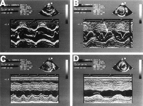
Figure 1 .
Typical M mode echocardiogram from a patient with hypertrophic cardiomyopathy highlighting the four main echocardiographic features of the condition. (A) Midsystolic closure of the aortic valve (arrowhead); (B) systolic anterior motion of the mitral valve (arrow) and asymmetric left ventricular hypertrophy together with a small, vigorously contracting left ventricle. In frames (C) and (D), the M mode beam passes through the septum and posterior wall beyond the mitral valve, at the level of the papillary muscles and apex, demonstrating the large reduction of left ventricular end systolic dimensions. Reproduced from Nihoyannopoulos and McKenna with permission of Churchill Livingstone.10

