Full Text
The Full Text of this article is available as a PDF (222.8 KB).
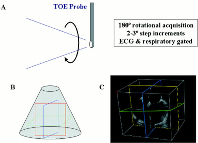
Figure 1 .
(A) Acquisition of multiple 2D images by rotation of multiplane transoesophageal probe through 180°. (B) Assimilation of multiple 2D imaging planes to generate a 3D volume of data. Coloured lines show typical cutplanes within the dataset. (C) Example of 3D dataset with demonstration of cutplanes.
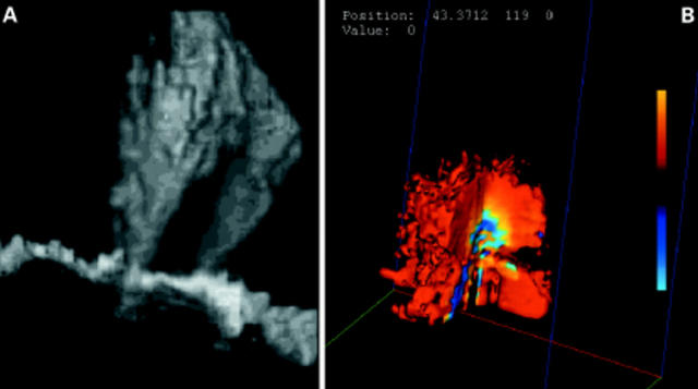
Figure 2 .
(A) Grey scale reconstruction of twin regurgitant jets extending upwards into the cavity of the left atrium. (B) Digital 3D colour Doppler rendering of a flow convergence region (in vitro acquisition).
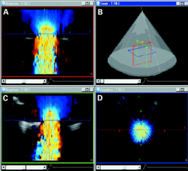
Figure 3 .
Localisation of the vena contracta in the 3D dataset (in vitro acquisition). Colour Doppler flow data have been acquired parallel to flow. (A) and (C) show direction of flow from top to bottom in the two. (D) A perpendicular (blue cutplane) cut through the narrowest portion of the regurgitant jet, the vena contracta.
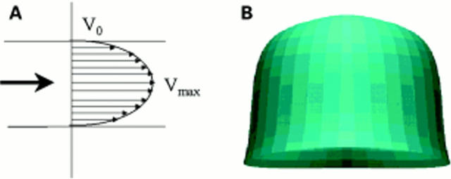
Figure 4 .
(A) Example of laminar flow pattern, such as might be encountered in the ventricular outflow tracts or the great vessels. (B) 3D reconstruction of a laminar flow profile (in vitro acquisition).
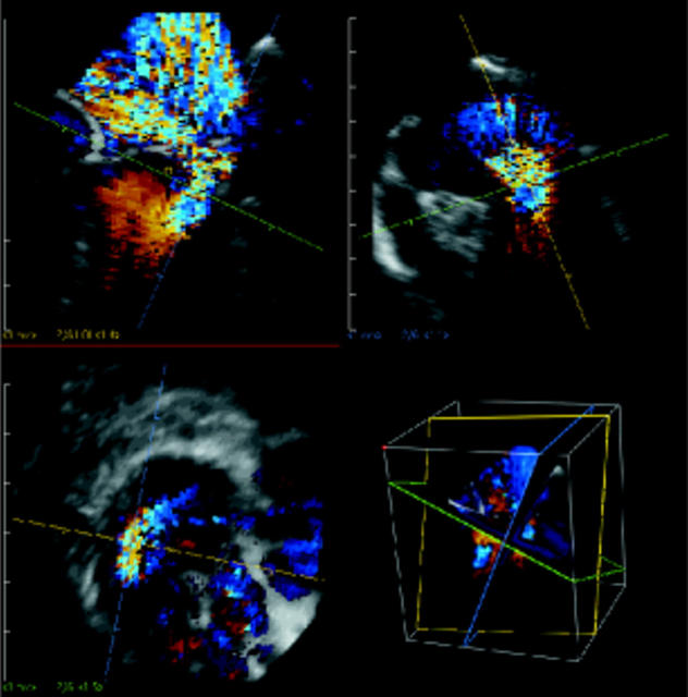
Figure 5 .
Identification of the vena contracta in a case of anterior mitral valve leaflet prolapse. In spite of the eccentricity of the jet, the cutplanes can still be manipulated to produce a cross section of the vena contracta (bottom left panel). It is then a simple process to measure its area. This should give an approximation of the regurgitant orifice area.
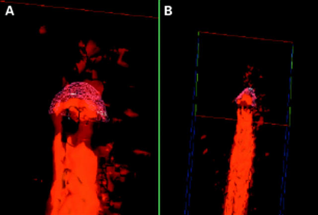
Figure 6 .
Calculation of the flow convergence region surface area in the 3D dataset. (A) is a magnified view of (B). A wire model framework has been fitted to the surface of the flow convergence region. Surface area computations are made using this model. Note that no geometric assumptions are involved in the measurement of the surface area.
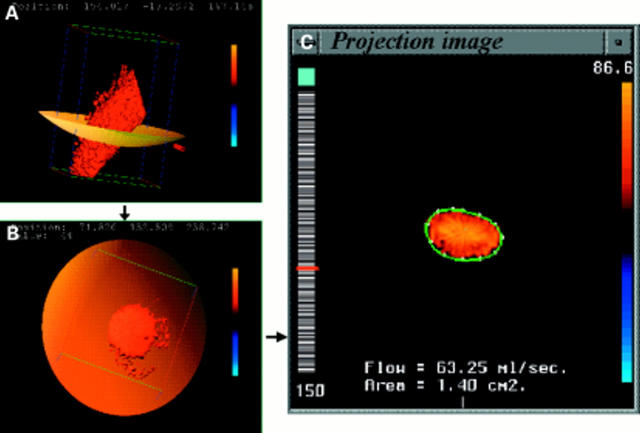
Figure 7 .
Computation of instantaneous flow rate from a digital 3D Doppler dataset, acquired from an in vitro tube model of laminar flow. The colour flow cross section is identified within the dataset (A), (B). The computer performs a spatial integration of the individual velocity vectors contained within the flow cross-section (C) to generate an instantaneous flow rate.
Selected References
These references are in PubMed. This may not be the complete list of references from this article.
- Aida S., Shiota T., Tsujino H., Sahn D. J. Quantification of aortic regurgitant volume by a newly developed automated cardiac flow measurement method: an in vitro study. J Am Soc Echocardiogr. 1998 Sep;11(9):874–881. doi: 10.1016/s0894-7317(98)70007-0. [DOI] [PubMed] [Google Scholar]
- Ascah K. J., Stewart W. J., Levine R. A., Weyman A. E. Doppler-echocardiographic assessment of cardiac output. Radiol Clin North Am. 1985 Dec;23(4):659–670. [PubMed] [Google Scholar]
- Bargiggia G. S., Tronconi L., Sahn D. J., Recusani F., Raisaro A., De Servi S., Valdes-Cruz L. M., Montemartini C. A new method for quantitation of mitral regurgitation based on color flow Doppler imaging of flow convergence proximal to regurgitant orifice. Circulation. 1991 Oct;84(4):1481–1489. doi: 10.1161/01.cir.84.4.1481. [DOI] [PubMed] [Google Scholar]
- Borrás X., Carreras F., Augé J. M., Pons-Lladó G. Prospective validation of detection and quantitative assessment of chronic aortic regurgitation by a combined echocardiographic and Doppler method. J Am Soc Echocardiogr. 1988 Nov-Dec;1(6):422–429. doi: 10.1016/s0894-7317(88)80024-5. [DOI] [PubMed] [Google Scholar]
- Dujardin K. S., Enriquez-Sarano M., Bailey K. R., Nishimura R. A., Seward J. B., Tajik A. J. Grading of mitral regurgitation by quantitative Doppler echocardiography: calibration by left ventricular angiography in routine clinical practice. Circulation. 1997 Nov 18;96(10):3409–3415. doi: 10.1161/01.cir.96.10.3409. [DOI] [PubMed] [Google Scholar]
- Fehske W., Omran H., Manz M., Köhler J., Hagendorff A., Lüderitz B. Color-coded Doppler imaging of the vena contracta as a basis for quantification of pure mitral regurgitation. Am J Cardiol. 1994 Feb 1;73(4):268–274. doi: 10.1016/0002-9149(94)90232-1. [DOI] [PubMed] [Google Scholar]
- Giesler M., Grossmann G., Schmidt A., Kochs M., Langhans J., Stauch M., Hombach V. Color Doppler echocardiographic determination of mitral regurgitant flow from the proximal velocity profile of the flow convergence region. Am J Cardiol. 1993 Jan 15;71(2):217–224. doi: 10.1016/0002-9149(93)90741-t. [DOI] [PubMed] [Google Scholar]
- Grayburn P. A., Fehske W., Omran H., Brickner M. E., Lüderitz B. Multiplane transesophageal echocardiographic assessment of mitral regurgitation by Doppler color flow mapping of the vena contracta. Am J Cardiol. 1994 Nov 1;74(9):912–917. doi: 10.1016/0002-9149(94)90585-1. [DOI] [PubMed] [Google Scholar]
- Helmcke F., Nanda N. C., Hsiung M. C., Soto B., Adey C. K., Goyal R. G., Gatewood R. P., Jr Color Doppler assessment of mitral regurgitation with orthogonal planes. Circulation. 1987 Jan;75(1):175–183. doi: 10.1161/01.cir.75.1.175. [DOI] [PubMed] [Google Scholar]
- Hozumi T., Yoshida K., Akasaka T., Takagi T., Yamamuro A., Yagi T., Yoshikawa J. Automated assessment of mitral regurgitant volume and regurgitant fraction by a newly developed digital color Doppler velocity profile integration method. Am J Cardiol. 1997 Nov 15;80(10):1325–1330. doi: 10.1016/s0002-9149(97)00673-5. [DOI] [PubMed] [Google Scholar]
- Irvine T., Derrick G., Morris D., Norton M., Kenny A. Three-dimensional echocardiographic reconstruction of mitral valve color Doppler flow events. Am J Cardiol. 1999 Nov 1;84(9):1103-6, A10. doi: 10.1016/s0002-9149(99)00512-3. [DOI] [PubMed] [Google Scholar]
- Ishii M., Jones M., Shiota T., Yamada I., Heinrich R. S., Holcomb S. R., Yoganathan A. P., Sahn D. J. Quantifying aortic regurgitation by using the color Doppler-imaged vena contracta: a chronic animal model study. Circulation. 1997 Sep 16;96(6):2009–2015. doi: 10.1161/01.cir.96.6.2009. [DOI] [PubMed] [Google Scholar]
- Jiang L., Siu S. C., Handschumacher M. D., Luis Guererro J., Vazquez de Prada J. A., King M. E., Picard M. H., Weyman A. E., Levine R. A. Three-dimensional echocardiography. In vivo validation for right ventricular volume and function. Circulation. 1994 May;89(5):2342–2350. doi: 10.1161/01.cir.89.5.2342. [DOI] [PubMed] [Google Scholar]
- Lee W., Rokey R., Cotton D. B. Noninvasive maternal stroke volume and cardiac output determinations by pulsed Doppler echocardiography. Am J Obstet Gynecol. 1988 Mar;158(3 Pt 1):505–510. doi: 10.1016/0002-9378(88)90014-2. [DOI] [PubMed] [Google Scholar]
- Miyatake K., Izumi S., Okamoto M., Kinoshita N., Asonuma H., Nakagawa H., Yamamoto K., Takamiya M., Sakakibara H., Nimura Y. Semiquantitative grading of severity of mitral regurgitation by real-time two-dimensional Doppler flow imaging technique. J Am Coll Cardiol. 1986 Jan;7(1):82–88. doi: 10.1016/s0735-1097(86)80263-7. [DOI] [PubMed] [Google Scholar]
- Mori Y., Shiota T., Jones M., Wanitkun S., Irvine T., Li X., Delabays A., Pandian N. G., Sahn D. J. Three-dimensional reconstruction of the color Doppler-imaged vena contracta for quantifying aortic regurgitation: studies in a chronic animal model. Circulation. 1999 Mar 30;99(12):1611–1617. doi: 10.1161/01.cir.99.12.1611. [DOI] [PubMed] [Google Scholar]
- Nosir Y. F., Stoker J., Kasprzak J. D., Lequin M. H., Dall'Agata A., Ten Cate F. J., Roelandt J. R. Paraplane analysis from precordial three-dimensional echocardiographic data sets for rapid and accurate quantification of left ventricular volume and function: a comparison with magnetic resonance imaging. Am Heart J. 1999 Jan;137(1):134–143. doi: 10.1016/s0002-8703(99)70469-2. [DOI] [PubMed] [Google Scholar]
- Papavassiliou D. P., Parks W. J., Hopkins K. L., Fyfe D. A. Three-dimensional echocardiographic measurement of right ventricular volume in children with congenital heart disease validated by magnetic resonance imaging. J Am Soc Echocardiogr. 1998 Aug;11(8):770–777. doi: 10.1016/s0894-7317(98)70051-3. [DOI] [PubMed] [Google Scholar]
- Pini R., Giannazzo G., Di Bari M., Innocenti F., Rega L., Casolo G., Devereux R. B. Transthoracic three-dimensional echocardiographic reconstruction of left and right ventricles: in vitro validation and comparison with magnetic resonance imaging. Am Heart J. 1997 Feb;133(2):221–229. doi: 10.1016/s0002-8703(97)70212-6. [DOI] [PubMed] [Google Scholar]
- Robson S. C., Murray A., Peart I., Heads A., Hunter S. Reproducibility of cardiac output measurement by cross sectional and Doppler echocardiography. Br Heart J. 1988 Jun;59(6):680–684. doi: 10.1136/hrt.59.6.680. [DOI] [PMC free article] [PubMed] [Google Scholar]
- Roelandt J. R. Three-dimensional echocardiography: new views from old windows. Br Heart J. 1995 Jul;74(1):4–6. doi: 10.1136/hrt.74.1.4. [DOI] [PMC free article] [PubMed] [Google Scholar]
- Roelandt J. R., Yao J., Kasprzak J. D. Three-dimensional echocardiography. Curr Opin Cardiol. 1998 Nov;13(6):386–396. doi: 10.1097/00001573-199811000-00002. [DOI] [PubMed] [Google Scholar]
- Shiota T., Jones M., Delabays A., Li X., Yamada I., Ishii M., Acar P., Holcomb S., Pandian N. G., Sahn D. J. Direct measurement of three-dimensionally reconstructed flow convergence surface area and regurgitant flow in aortic regurgitation: in vitro and chronic animal model studies. Circulation. 1997 Nov 18;96(10):3687–3695. doi: 10.1161/01.cir.96.10.3687. [DOI] [PubMed] [Google Scholar]
- Shiota T., Jones M., Yamada I., Heinrich R. S., Ishii M., Sinclair B., Holcomb S., Yoganathan A. P., Sahn D. J. Effective regurgitant orifice area by the color Doppler flow convergence method for evaluating the severity of chronic aortic regurgitation. An animal study. Circulation. 1996 Feb 1;93(3):594–602. doi: 10.1161/01.cir.93.3.594. [DOI] [PubMed] [Google Scholar]
- Shiota T., Sinclair B., Ishii M., Zhou X., Ge S., Teien D. E., Gharib M., Sahn D. J. Three-dimensional reconstruction of color Doppler flow convergence regions and regurgitant jets: an in vitro quantitative study. J Am Coll Cardiol. 1996 May;27(6):1511–1518. doi: 10.1016/0735-1097(96)00009-5. [DOI] [PubMed] [Google Scholar]
- Spain M. G., Smith M. D., Grayburn P. A., Harlamert E. A., DeMaria A. N. Quantitative assessment of mitral regurgitation by Doppler color flow imaging: angiographic and hemodynamic correlations. J Am Coll Cardiol. 1989 Mar 1;13(3):585–590. doi: 10.1016/0735-1097(89)90597-4. [DOI] [PubMed] [Google Scholar]
- Stein P. D., Sabbah H. N. Nature of flow in large arteries. Monogr Atheroscler. 1990;15:54–62. [PubMed] [Google Scholar]
- Sun J. P., Pu M., Fouad F. M., Christian R., Stewart W. J., Thomas J. D. Automated cardiac output measurement by spatiotemporal integration of color Doppler data. In vitro and clinical validation. Circulation. 1997 Feb 18;95(4):932–939. doi: 10.1161/01.cir.95.4.932. [DOI] [PubMed] [Google Scholar]
- Van Camp G., Carlier S., Cosyns B., Plein D., Menassel M., Josse T., Verdonck P., Segers P., Vandenbossche J. L. Quantification of mitral regurgitation by the automated cardiac output method: an in vitro and in vivo study. J Am Soc Echocardiogr. 1998 Jun;11(6):643–651. doi: 10.1016/s0894-7317(98)70041-0. [DOI] [PubMed] [Google Scholar]
- Yao J., Masani N. D., Cao Q. L., Nikuta P., Pandian N. G. Clinical application of transthoracic volume-rendered three-dimensional echocardiography in the assessment of mitral regurgitation. Am J Cardiol. 1998 Jul 15;82(2):189–196. doi: 10.1016/s0002-9149(98)00305-1. [DOI] [PubMed] [Google Scholar]
- Yoganathan A. P., Cape E. G., Sung H. W., Williams F. P., Jimoh A. Review of hydrodynamic principles for the cardiologist: applications to the study of blood flow and jets by imaging techniques. J Am Coll Cardiol. 1988 Nov;12(5):1344–1353. doi: 10.1016/0735-1097(88)92620-4. [DOI] [PubMed] [Google Scholar]
- Zhou Y. Q., Faerestrand S., Matre K., Birkeland S. Velocity distributions in the left ventricular outflow tract and the aortic anulus measured with Doppler colour flow mapping in normal subjects. Eur Heart J. 1993 Sep;14(9):1179–1188. doi: 10.1093/eurheartj/14.9.1179. [DOI] [PubMed] [Google Scholar]


