Abstract
Despite the advent of sophisticated imaging systems, plain radiography continues to be a valuable outcome variable in clinical trials of inflammatory disorders for a number of reasons. This paper discusses the pros and cons of the different ways in which radiographic data in trials is presented; the minimum time needed to demonstrate radiographic progression in the context of a clinical trial; and the best ways to statistically analyse radiographic data.
Full Text
The Full Text of this article is available as a PDF (125.8 KB).
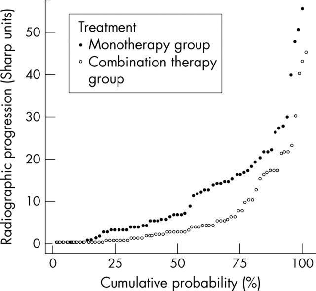
Figure 1.
Probability plots representing one year radiographic progression in both groups of the COBRA study (see table 1 footnote for details). Every symbol represents the score of an individual, and all scores are plotted against their cumulative probability.
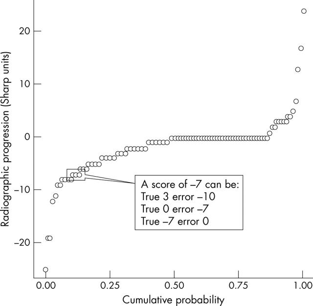
Figure 2.
Probability plot of an imaginary progression scenario. Negative progression scores do not necessarily imply repair. Every individual score represents a combination of true change and measurement error. It is impossible to distinguish both at the individual patient level.
Figure 3.

(A) Probability plots of progression scores obtained from a three month clinical trial, in which sets of radiographs were scored with known time order (chronological; red) and with unknown time order (paired; blue). (B, C) Probability plots of progression scores obtained from a three month clinical trial in which sets of radiographs were scored with known time order (chronological; B) and with unknown time order (paired; C), stratified for the presence (blue) or absence (red) of radiographic damage at baseline.
Selected References
These references are in PubMed. This may not be the complete list of references from this article.
- Boers M., Verhoeven A. C., Markusse H. M., van de Laar M. A., Westhovens R., van Denderen J. C., van Zeben D., Dijkmans B. A., Peeters A. J., Jacobs P. Randomised comparison of combined step-down prednisolone, methotrexate and sulphasalazine with sulphasalazine alone in early rheumatoid arthritis. Lancet. 1997 Aug 2;350(9074):309–318. doi: 10.1016/S0140-6736(97)01300-7. [DOI] [PubMed] [Google Scholar]
- Bruynesteyn K., Landewé R., van der Linden Sj, van der Heijde D. Radiography as primary outcome in rheumatoid arthritis: acceptable sample sizes for trials with 3 months' follow up. Ann Rheum Dis. 2004 Mar 22;63(11):1413–1418. doi: 10.1136/ard.2003.014043. [DOI] [PMC free article] [PubMed] [Google Scholar]
- Bruynesteyn Karin, Van Der Heijde Désirée, Boers Maarten, Saudan Ariane, Peloso Paul, Paulus Harold, Houben Harry, Griffiths Bridget, Edmonds John, Bresnihan Barry. Detecting radiological changes in rheumatoid arthritis that are considered important by clinical experts: influence of reading with or without known sequence. J Rheumatol. 2002 Nov;29(11):2306–2312. [PubMed] [Google Scholar]
- Klareskog Lars, van der Heijde Désirée, de Jager Julien P., Gough Andrew, Kalden Joachim, Malaise Michel, Martín Mola Emilio, Pavelka Karel, Sany Jacques, Settas Lucas. Therapeutic effect of the combination of etanercept and methotrexate compared with each treatment alone in patients with rheumatoid arthritis: double-blind randomised controlled trial. Lancet. 2004 Feb 28;363(9410):675–681. doi: 10.1016/S0140-6736(04)15640-7. [DOI] [PubMed] [Google Scholar]
- Landewé Robert B. M., Boers Maarten, Verhoeven Arco C., Westhovens Rene, van de Laar Mart A. F. J., Markusse Harry M., van Denderen J. Christiaan, Westedt Marie Louise, Peeters Andre J., Dijkmans Ben A. C. COBRA combination therapy in patients with early rheumatoid arthritis: long-term structural benefits of a brief intervention. Arthritis Rheum. 2002 Feb;46(2):347–356. doi: 10.1002/art.10083. [DOI] [PubMed] [Google Scholar]
- Landewé Robert, van der Heijde Désirée. Radiographic progression depicted by probability plots: presenting data with optimal use of individual values. Arthritis Rheum. 2004 Mar;50(3):699–706. doi: 10.1002/art.20204. [DOI] [PubMed] [Google Scholar]
- Plant M. J., Jones P. W., Saklatvala J., Ollier W. E., Dawes P. T. Patterns of radiological progression in early rheumatoid arthritis: results of an 8 year prospective study. J Rheumatol. 1998 Mar;25(3):417–426. [PubMed] [Google Scholar]
- Welsing P. M., van Gestel A. M., Swinkels H. L., Kiemeney L. A., van Riel P. L. The relationship between disease activity, joint destruction, and functional capacity over the course of rheumatoid arthritis. Arthritis Rheum. 2001 Sep;44(9):2009–2017. doi: 10.1002/1529-0131(200109)44:9<2009::AID-ART349>3.0.CO;2-L. [DOI] [PubMed] [Google Scholar]
- Welsing Paco M. J., Landewé Robert B. M., van Riel Piet L. C. M., Boers Maarten, van Gestel Anke M., van der Linden Sjef, Swinkels Hilde L., van der Heijde Désirée M. F. M. The relationship between disease activity and radiologic progression in patients with rheumatoid arthritis: a longitudinal analysis. Arthritis Rheum. 2004 Jul;50(7):2082–2093. doi: 10.1002/art.20350. [DOI] [PubMed] [Google Scholar]
- Zeger S. L., Liang K. Y. Longitudinal data analysis for discrete and continuous outcomes. Biometrics. 1986 Mar;42(1):121–130. [PubMed] [Google Scholar]
- van der Heijde D., Landewé R. Imaging: do erosions heal? Ann Rheum Dis. 2003 Nov;62 (Suppl 2):ii10–ii12. doi: 10.1136/ard.62.suppl_2.ii10. [DOI] [PMC free article] [PubMed] [Google Scholar]
- van der Heijde Désirée, Landewé Robert, Klareskog Lars, Rodríguez-Valverde Vicente, Settas Lucas, Pedersen Ronald, Fatenejad Saeed. Presentation and analysis of data on radiographic outcome in clinical trials: experience from the TEMPO study. Arthritis Rheum. 2005 Jan;52(1):49–60. doi: 10.1002/art.20775. [DOI] [PubMed] [Google Scholar]


