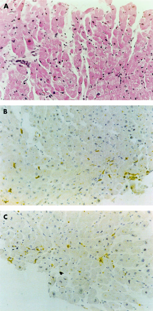
Figure 2.
Borderline myocarditis. (A) Haematoxylin and eosin staining shows absence of myocardial necrosis and a few inflammatory cell infiltrates. Immunohistochemical staining for (B) CD43 (T leucocytes) and (C) CD68 (macrophages) shows the presence of leucocytes within the myocardium. Original magnification ×180.

