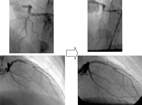
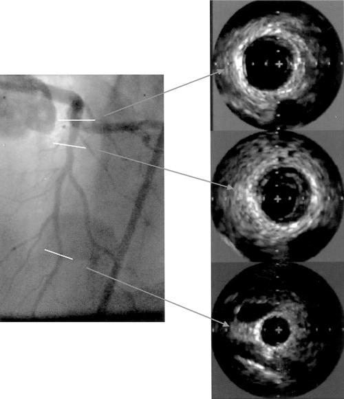
A 23 year old male body builder presented with a recent onset of central chest pain. He was a smoker with cholesterol concentration of 7.l mmol/l on admission. He had been taking anabolic steroids (methandrostenelone 20 mg daily) for three months. Anteroseptal T wave inversion on a 12 lead ECG along with elevated troponin T prompted early coronary angiography. This revealed multiple filling defects in the mid left anterior descending (LAD) artery consistent with the presence of thrombus (below left). His LAD filled by collaterals from the right coronary artery. The rest of his coronary arteries were smooth and unobstructed. Repeat angiography was performed 48 hours later after treatment with abciximab, aspirin, and low molecular weight heparin and revealed complete dissolution of all thrombus. Intravascular ultrasound (IVUS) examination performed at this time revealed a segment of eccentric atheroma at the site of the previous filling defects (below right); the echogenicity was uniform with no focal echo lucent regions. The remainder of the LAD was normal at IVUS examination. The patient made an uneventful recovery and was discharged well to follow up.
Figure 1.
Top and bottom left: left anterior oblique and right anterior oblique views of left coronary artery with filling defect. Top and bottom right: same views after 48 hours of antithrombotic treatment (heparin, aspirin, and abciximab).
There have been several case reports of acute myocardial infarction in young male athletes using anabolic steroids. The mechanism is unclear but may involve the adverse effects on thrombosis and lipid profile. Some reports suggest thrombosis in “normal” coronary arteries, but underlying atheroma cannot be excluded without IVUS. This case supports the concept that both atheroma and the thrombogenic effects of anabolic steroids may be necessary for vessel occlusion.
Figure 2.
Intravascular ultrasound images at designated sites of the left anterior descending artery after 48 hours of antithrombotic treatment


