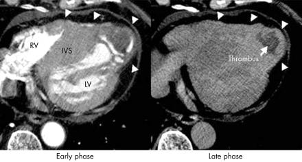
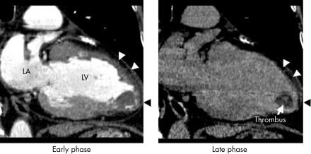
A 65 year old man with hypertrophic cardiomyopathy presented at our hospital because of dyspnoea on effort. An echocardiogram revealed a very large hypertrophy of the interventricular septum (IVS) with reduced apical wall motion and thinning of the apical wall of the left ventricle (LV), but a thrombus could not be observed in the LV. Therefore, we suspected the dilated form of hypertrophic cardiomyopathy. To evaluate the characteristics of the myocardium and to determine if a thrombus existed in the LV, ECG gated enhanced multislice computed tomography (CT) (Light Speed Ultra, General Electric, Milwaukee, Wisconsin, USA) was performed with a 1.25 mm slice thickness, helical pitch 3.25. Following intravenous injection of 100 ml of iodinated contrast material (350 mgI/ml), CT scanning was performed with retrospective ECG gated reconstruction at 30 seconds and eight minutes after the injection, and volume data were extracted from end diastole. In the axial source images and multiplanar reconstruction images representing the long axis of the LV, a thrombus and thinning of the wall in the apex of the LV (arrowheads) could be observed with a huge hypertrophy of IVS in the early phase. In the late phase, the thinned wall of the apical portion of the LV and the apical wall of the right ventricle (RV) were abnormally enhanced, compared with the huge, hypertrophic IVS, suggesting that they had caused fibrotic change. Since the reduced apical motion of the LV identified by the echocardiogram, caused by fibrotic change identified by CT, might produce large thrombi, anticoagulant treatment was initiated.
. 2003 Aug;89(8):858. doi: 10.1136/heart.89.8.858
Thinned myocardial fibrosis with thrombus in the dilated form of hypertrophic cardiomyopathy demonstrated by multislice computed tomography
N Funabashi
, K Yoshida
, I Komuro
N Funabashi
Find articles by N Funabashi
K Yoshida
Find articles by K Yoshida
I Komuro
Find articles by I Komuro
komuro-tky@umin.ac.jp
Copyright © Copyright 2003 by Heart
PMCID: PMC1767795 PMID: 12860857


