An 18 year old man presented with progressive dyspnoea for three months. Seven months before admission he had suffered from choreatic movements of the right arm. A month before admission the general practitioner treated him for diagnosed pericarditis with high dosages of salicylates.
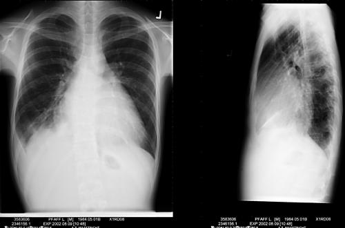
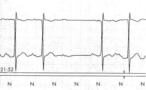
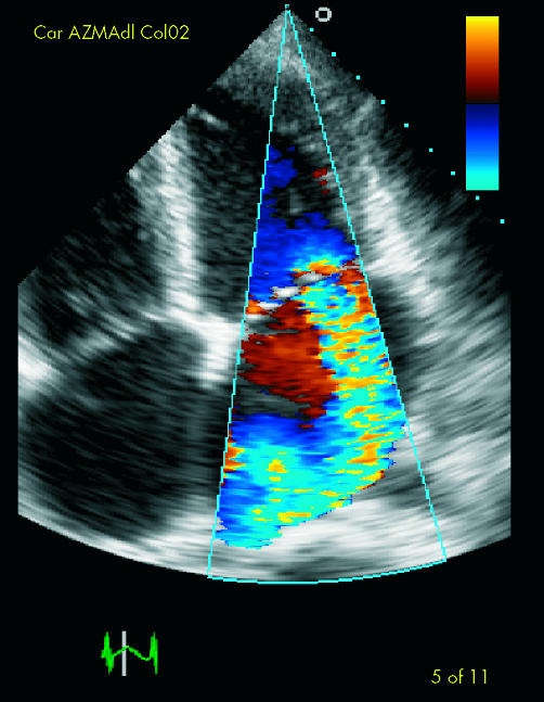
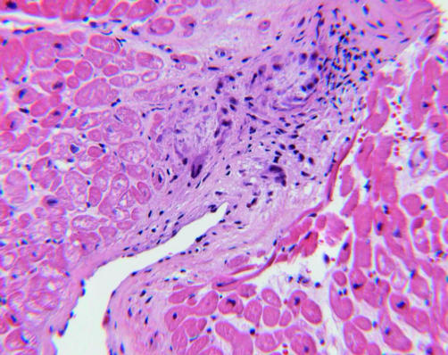
At physical examination the patient appeared chronically ill and in biventricular congestive cardiac failure, with findings of mild aortic regurgitation (AR), severe mitral (MR) and tricuspid regurgitation (TR), in the presence of a dilated left ventricle. The chest x ray showed cardiomegaly with pronounced left atrial dilatation (below, upper panel). The 12 lead ECG showed a sinus tachycardia, first degree atrioventricular conduction delay, and left atrial dilatation, while rhythm monitoring showed intermittent second degree type II atrioventricular conduction delay (below, lower panel). Laboratory assessment revealed a raised C reactive protein, erythrocyte sedimentation rate, anti-streptolysin O titre, and anti-DNAse B. The culture of the throat swab remained negative. Transthoracic echocardiography (right, upper panel) confirmed mild AR, and severe MR and TR with a mildly dilated left ventricle and preserved systolic function. The right ventricular pressures were raised (50 mm Hg). The clinical diagnosis of rheumatic carditis was considered in the presence of two major and two minor revised Jones criteria. Since thiscondition is rarely diagnosed in the Netherlands, and diagnostic uncertainty remained, it was decided to obtain left ventricular myocardial biopsies. In two of the three samples Aschoff bodies, pathognomonic for rheumatic carditis, were found (right, lower panel).
The patient was treated with penicillin, loop diuretics, angiotensin converting enzyme inhibitors, digitalis, and β blockers and his condition improved significantly.


