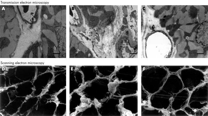
Figure 2.
Transmission and scanning electron micrographs. The LETO rats showed normal appearances (A and D). In the OLETF rats, thickened capillary basement membranes and interstitial fibrosis were prominent (B and E). All the changes were smaller in the OLETF+ARB subgroup (C and F). Scale bar = 1 μm. ARB, angiotensin II receptor blockade.

