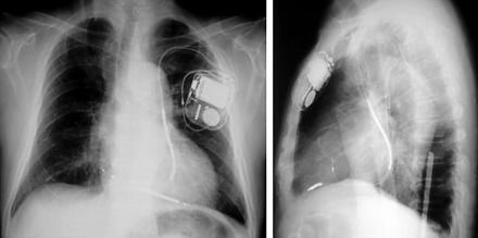
A 67 year old man with chronic renal failure, hypertension and ischaemic heart disease was admitted for polymorphic ventricular tachycardia with syncope. Echocardiography revealed left ventricular hypertrophy with moderately impaired function. Amiodarone was administered for these arrhythmias, but recurrent episodes were experienced during haemodialysis. Therefore, an implantable cardioverter-defibrillator (ICD) was implanted at the left anterior chest on the other side of the shunt vein. Although the left cephalic vein was too small for insertion of a ventricular lead, an atrial lead was inserted from there, and the ventricular lead was inserted from the left subclavian vein. During the procedure, a persistent left superior vena cava (PLSVC) was detected incidentally, as both leads ran through an unusual left sided downward course. The Medtronic model 6945 (65 cm) screw-in ventricular lead was successfully affixed to the right ventricular apex by forming the stylet into a U shape in the right atrium. The Intermedics model 438-35S (52 cm) atrial lead was fixated in the right atrium (see panels). Atrial and ventricular sensing and stimulation thresholds were acceptable. Ventricular fibrillation induced by a T wave shock was successfully defibrillated twice with 20 joules of energy.
PLSVC is present in approximately 0.5% of the population, and a transvenous pacemaker or ICD implantation is sometimes difficult or even impossible in those cases. If transvenous implantation via PLSVC is intended, it is recommended to use a screw-in type electrode with sufficient length so that a loop in the right atrium can be formed for appropriate fixation to the right ventricle.


