Abstract
A 59 year old man undergoing investigation for chest pain was found at elective coronary angiography to have a single coronary artery; the left coronary had a normal distribution, with the right coronary originating as a continuation of the atrioventricular circumflex. His 30 year old daughter was admitted for elective coronary angiography for further investigation of a dilated cardiomyopathy. She was also found to have a single coronary artery. However, in her case, the right and left coronary arteries arose from the right sinus of Valsalva; the right coronary had a normal distribution, the left coronary passed anterior to the pulmonary trunk and aorta.
Keywords: congenital heart disease, coronary anomaly, single coronary artery
Congenital coronary anomalies occur rarely, varying from 0.3–1.3% of the population.1–5 Most such anomalies are of no clinical significance and are found incidentally at coronary angiography or at necropsy. We present an unusual case of a father and daughter, both of whom had a single coronary artery. To our knowledge no association of this nature has been previously described.
CASE REPORTS
Case 1
A 59 year old man was admitted for coronary angiography. He had a four year history of exertional angina, and stress testing was positive for ischaemia at a low level of exercise.
At cardiac catheterisation a single coronary artery was found, with the left coronary artery arising from the left sinus of Valsalva and the right coronary originating as a continuation of the atrioventricular circumflex artery. There was significant disease in both the left anterior descending artery and the left circumflex artery. The left ventricle was not dilated and systolic function was normal with an ejection fraction of 60%.
Subsequently, he underwent coronary artery bypass graft surgery.
Case 2
A 30 year old woman was admitted for further investigation. She had a history of hypertension treated with atenolol. During her second pregnancy she had noted dyspnoea, atypical chest pain, palpitations, and peripheral oedema.
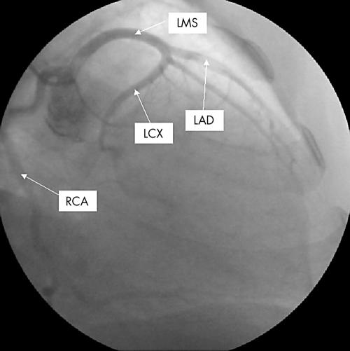
An echocardiogram was performed, which showed a mildly dilated left ventricle with moderate systolic dysfunction. This did not improve in the postpartum period. At cardiac catheterisation, a single coronary artery was found with both left and right coronary arteries arising from the right sinus of Valsalva (fig 1). Computed tomography confirmed that the left mainstem artery passed anterior to the pulmonary artery.
Figure 1.
Coronary angiogram of case 2, showing a single coronary artery with both left and right coronary arteries arising from the right sinus of Valsalva. LAD, left anterior descending artery; LCX, left circumflex artery, LMS, left main stem, RCA, right coronary artery.
DISCUSSION
Coronary anomalies are uncommon, but with the widespread availability of coronary angiography they are likely to be recognised more frequently. However, familial clustering of single coronary anomalies is extremely uncommon. A search of the literature in PubMed and Medline with the keywords “coronary anomaly”, “single coronary artery”, and “familial” found only one other report of a familial association of coronary anomalies. We have described a father and daughter who were both found to have a left circumflex artery arising from the right aortic sinus.6
In the first case the right coronary artery arises as a continuation of the left circumflex artery. This has previously been described in the literature, although it is one of the least common coronary anomalies with the highest reported incidence of only 0.035%.7,8
In the second case the left coronary artery shares a common origin with the right coronary artery arising from the right sinus of Valsalva, and therefore can be regarded as a type of single coronary artery. This has a reported incidence of 0.017%.3 Obtaining evidence of such a coronary anomaly is clinically important and is dependent on the anatomical subtype. Anomalies are classified according to the relation of the coronary anomaly to the aorta and pulmonary artery—that is, anterior, between, septal, posterior, and combined. Previous investigation of coronary anomalies and analysis of the causes of sudden cardiac death has identified the course of the left main coronary artery between the aorta and pulmonary artery as a potential cause of significant coronary ischaemia.3,9 This anomaly can present with exertional angina, dyspnoea, and palpitations, as well as sudden death. There are several proposed potential mechanisms for the clinical manifestation of this anomaly, such as compression of the left coronary artery between the aorta and pulmonary artery during exercise when the vessels become enlarged; and compromise of the lumen of the left coronary artery due to the acute angle formed at its origin from the right aortic sinus resulting in a slit-like orifice. The proposed management of this subgroup of patients is surgical because of the high incidence of sudden cardiac death, especially in the young (< 20 years), who seldom have associated coronary artery disease or other abnormalities.
We have presented an interesting association of different coronary anomalies in a family setting. Is there any evidence of an inherited or genetic link? Inheritance of different patterns of anomalous coronary circulation has not been extensively investigated; however, there is some evidence for a genetic effect on different phenotypic patterns of coronary arteries. This includes work showing similar patterns of circulation within the same family, as well as differences between races. Animal studies have also suggested a polygenic pattern of inheritance.10
The likelihood of finding such rare coronary circulations in two members of a family by chance alone is extremely small.
Supplementary Material
REFERENCES
- 1.Pillai SB, Khan MM, Diamond A, et al. The prevalence and types of coronary artery anomalies in Northern Ireland. Ulster Med J 2000;69:19–22. [PMC free article] [PubMed] [Google Scholar]
- 2.Click RL, Holmes DR, Vlietstra RE, et al. Anomalous coronary arteries: location, degree of atherosclerosis and effect on survival: a report from the coronary artery surgery study. J Am Coll Cardiol 1989;13:531–7. [DOI] [PubMed] [Google Scholar]
- 3.Yamanaka O, Hobbs RE. Coronary artery anomalies in 126,595 patients undergoing coronary arteriography. Cathet Cardiovasc Diagn 1990;21:28–40. [DOI] [PubMed] [Google Scholar]
- 4.Cieslinski G, Rapprich B, Kober G. Coronary anomalies: incidence and importance. Clin Cardiol 1993;16:711–5. [DOI] [PubMed] [Google Scholar]
- 5.Kimbiris D, Iskandrian AS, Segal BL, et al. Anomalous aortic origin of coronary arteries. Circulation 1978;58:606–14. [DOI] [PubMed] [Google Scholar]
- 6.Rowe L, Carmody TJ, Askenazi J. Anomalous origin of the left circumflex coronary artery from the right aortic sinus: a familial clustering. Cathet Cardiovasc Diagn 1993;29:277–8. [DOI] [PubMed] [Google Scholar]
- 7.Neuhaus R, Kober G. Single coronary artery with branching of the right coronary artery from the left atrioventricular ramus of the circumflex artery. Incidence and significance. Z Kardiol 1993;82:813–7. [PubMed] [Google Scholar]
- 8.Sheth M, Dovnarsky M, Cha SD, et al. Single coronary artery: right coronary artery originating from the distal left circumflex. Cathet Cardiovasc Diagn 1988;14:180–1. [DOI] [PubMed] [Google Scholar]
- 9.Roberts WC. Major anomalies of coronary arterial origin seen in adulthood. Am Heart J 1986;111:941–63. [DOI] [PubMed] [Google Scholar]
- 10.Bloor CM, Leon AS. The genetic determination of coronary artery patterns: a possible factor in atherogenesis. Ann N Y Acad Sci 1968;149:860–4. [DOI] [PubMed] [Google Scholar]
Associated Data
This section collects any data citations, data availability statements, or supplementary materials included in this article.


