Non-compaction of the left ventricle is a rare congenital cardiomyopathy characterised by a loosened spongy myocardium. The disorder may be associated with facial dysmorphism and familial recurrence and is caused by intrauterine arrest of compaction of the loose interwoven meshwork.
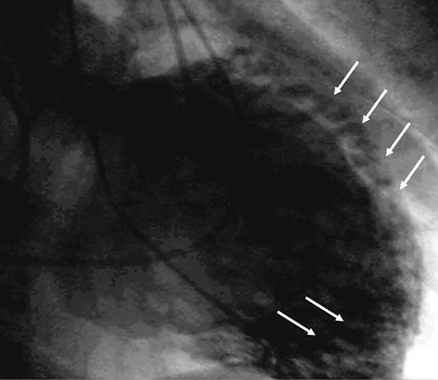
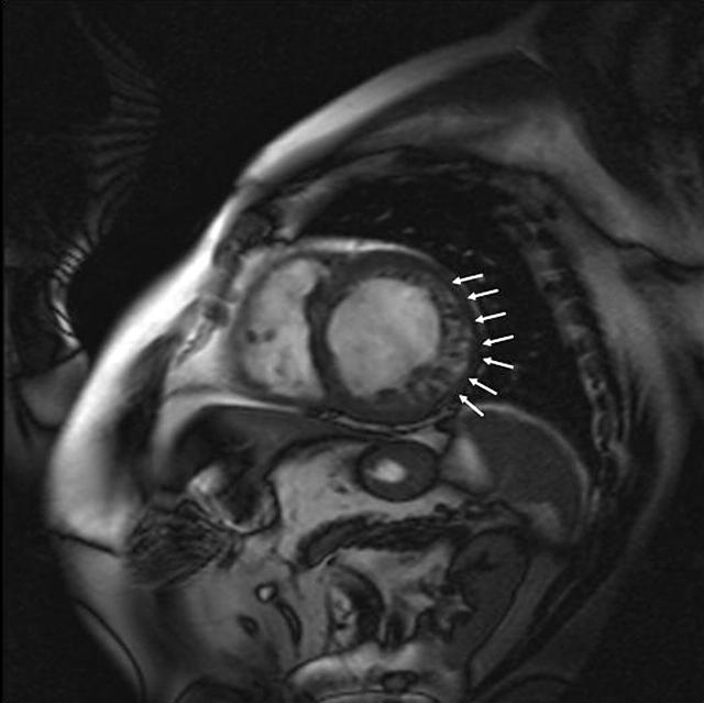
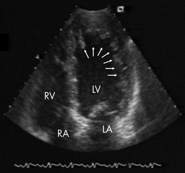
Left ventricular angiography (below left), cardiac magnetic resonance imaging (MRI) (below centre), and echocardiography (below right) typically reveal a dilated hypocontractile left ventricle with a two tailored wall; the inner zones of heavily spongious, trabecularised endocardial layers with deep intertrabecular recesses can be distinguished from thin outer zones of compacted myocardium. Histological examination confirms the spongy appearance with deep intertrabecular recesses, lined by endothelium, which spread close to the epicardial surface.
Prompt recognition of the disease is mandatory because of its high mortality and morbidity. Heart failure, thromboembolic events, and ventricular arrhythmias all have been reported. Besides familial screening and risk stratification, treatment is directed to prevent and manage heart failure, ventricular arrhythmias, and cardiac thromboembolism.
Figure 1.
Left ventricular angiography, 30° right anterior oblique projection. Arrow indicates non-compacted myocardium.
Figure 2.
MRI double inversion recovery images in diastole in left ventricular short axis view. Arrows indicate thickened non-compacted myocardial wall.
Figure 3.
Echo apical long axis view. Arrows indicate non-compacted left ventricle. LV, left ventricle; RV, right ventricle; LA, left atrium; RA, right atrium.


