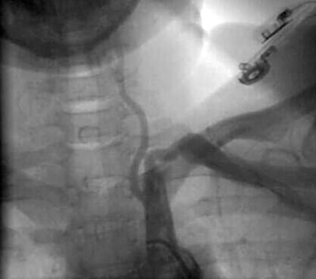
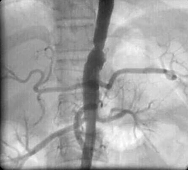
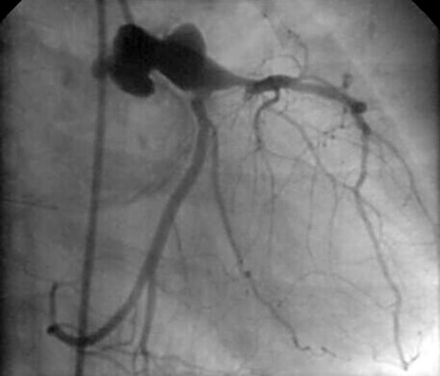
A 30 year old women was admitted to our hospital for evaluation of syncope. Her blood pressure was 100/70 mm Hg in the left arm, while it was not recordable in the right arm. Systolic bruit was present over the right supraclavicular region. There was a blowing diastolic murmur over the left sternal border. ECG was normal. Laboratory studies gave either negative or normal results. There was no evidence of systemic inflammation. Erythrocyte sedimentation rate was normal, C reactive protein was negative, and blood counts were normal. Additionally rheumatoid factor and ANA were negative. She underwent angiographic evaluation with provisional diagnosis of Takayasu arteritis (TA). Aortogram revealed mild aortic regurgitation, total occlusion of the right subclavian artery, narrowing of the proximal left subclavian artery (below, left panel), and stenosis of the supra renal aorta (middle panel) consistent with a diagnosis of TA. Coronary angiogram showed a large aneurysm of the left main coronary artery (right panel).
Most of the discrete coronary artery aneurysms are atherosclerotic in origin; other causes of coronary aneurysms include Kawasaki disease, Takayasu disease, polyarteritis nodosa, and systemic lupus erythematosis. TA is a chronic inflammatory vasculitis characterised by stenosis or obliteration of large and medium sized arteries. Morbidity results from arterial stenosis and organ ischaemia as well as from aneurysm formation. The cardiac sequelae of TA are far more commonly due to aortic regurgitation and inadequately treated hypertension than arteritis affecting the coronary vessels. Coronary arteries are affected in approximately 10% of the cases. Most of the lesions cause luminal narrowing; however, coronary aneurysm formation is extremely rare. The left main coronary artery is the least frequently involved artery. Aneurysm formation results from extensive destruction of elastic fibres in the media of the involved arterial wall as well as from arterial hypertension. A coronary aneurysm often causes stasis of blood flow and results in mural thrombus and myocardial infarction; saccular aneurysm may get thrombosed, ruptured or enlarged, resulting in myocardial infarction, cardiac tamponade or sudden death.
Figure 1.
Left subclavian angiogram showing the characteristic narrowing in the subclavian artery.
Figure 2.
Aortogram showing narrowing of the supra renal aorta.
Figure 3.
Coronary angiogram in the right anterior oblique showing aneurysm of the left main coronary artery.


