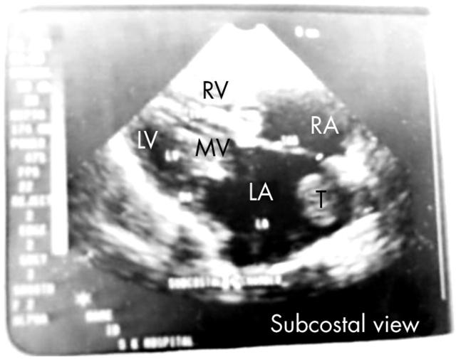
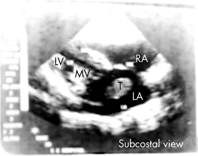
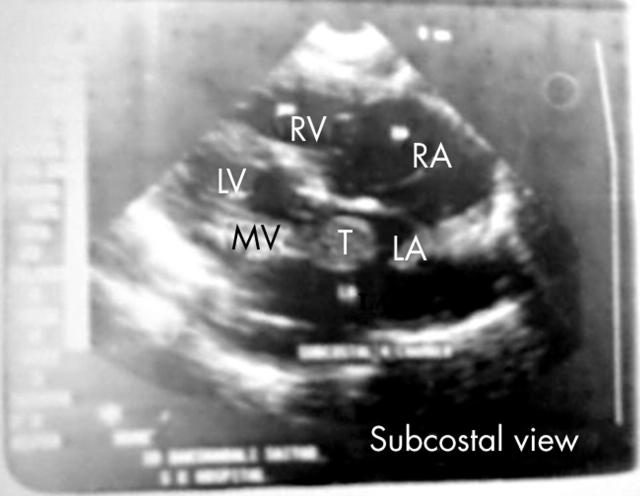
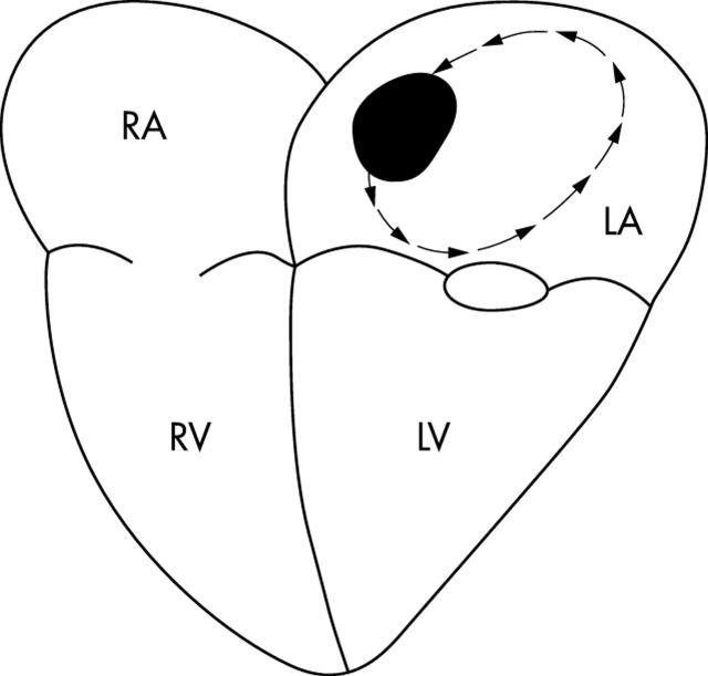
A 78 year old male patient presented with dyspnoea (New York Heart Association functional class III) with past history of left sided hemiparesis two years previously with atrial fibrillation on ECG. Chest x ray showed evidence of enlargement of the left atrium, right ventricle, and right atrium with redistribution of blood flow toward the lung apices.
M mode echocardiography showed multiple dense lines over the mitral valve area (thickened valve), reduced E-F slope of mitral valve (15 mm/s), with erratically moving multiple lines in an enlarged left atrium (73 mm).
Two dimensional echocardiography showed thickened, non-pliable leaflets, and a calcified valve annulus with a mitral valve area of approximately 0.7 cm2.
A globular, hyperechoic mass (thrombus) of 3 × 2 cm was detected in an enlarged left atrium. It was moving freely from the upper part of the left atrium to the lower part and was bouncing back after striking the mitral valve leaflets. The mass was unable to find its way through the mitral valve as it was larger than the mitral valve orifice. The mass was a dislodged thrombus that had failed to embolise.


