An asymptomatic 29 year old woman with left ventricular aneurysms was referred to our institution for surgical treatment. At the age of 1 year she had undergone closure of a membranous ventricular septal defect at another institution. The defect was closed with a synthetic patch through a combined right transatrial–transventricular approach.
Four years later, during hospitalisation for pneumonia, a chest x ray revealed cardiomegaly (cardiothoracic index 0.65). Echocardiography and subsequent cardiac angiography revealed the presence of two aneurysms on the left ventricle, one on the anterior wall and another on the posterior wall.
Two dimensional echocardiography showed a dyskinetic wall motion at the level of the aneurysms, while left ventricular function was within normal limits (ejection fraction 48%).
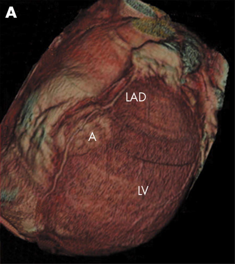
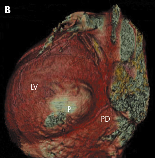
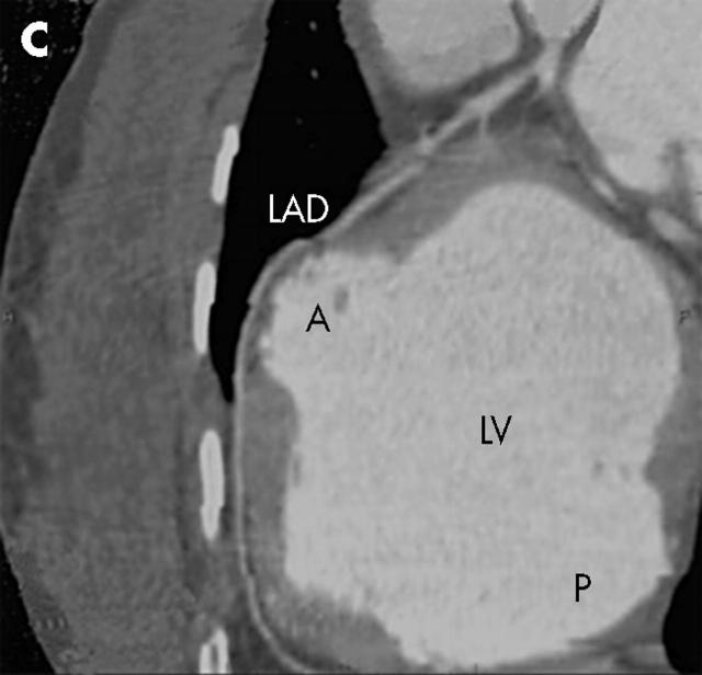
A multislice computed tomographic (CT) scan of the heart with three dimensional reconstruction allowed us to define the external anatomy of the two aneurysms and their relation with the left anterior descending and posterior descending coronary arteries (panels A–C: A, anterior aneurysm; LAD, left anterior descending coronary artery; LV, left ventricle; P, posterior aneurysm; PD, posterior descending coronary artery).
Multislice CT scanning of the heart with three dimensional reconstruction represents a reliable non-invasive method to assess the size and landmarks of left ventricular aneurysms and their relation to other heart structures.
Figure 1.


