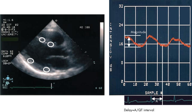
Figure 2.
Parasternal long axis integrated backscatter (IB) imaging interfaced with acoustic densitometry (left) and a representative curve of cycle dependent variation of myocardial IB (CV-IB) (right). An elliptical region of interest was placed in the mid or basal myocardial segment of the interventricular septum and posterior wall.

