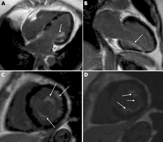
A 53 year old woman with Churg-Strauss syndrome and a history of asthma, pulmonary hyper-eosinophilia, and epileptic seizures was admitted to hospital with atypical chest pains. Her ECG showed generalised T wave inversions accompanied by TnI rise up to 7.8 μmol/l and hyper-eosinophilia (white blood count 28.8 × 10⁹/l, with 60% eosinophils, antineutrophil cytoplasmic antibodies (ANCA), and auto-antibodies were negative. Her symptoms and eosinophil counts responded well to steroid treatment. Subsequently, she was referred for rest and dobutamine stress echocardiography (DSE) to assess myocardial involvement and inducible myocardial ischaemia. The rest echocardiogram raised the suspicion of cardiac involvement, showing echogenic deposits in the basal septum; however, the DSE revealed no inducible ischaemia. For further clarification of myocardial involvement the patient was referred for cardiovascular magnetic resonance (CMR).
Contrast enhanced CMR 10–20 minutes after injection of 0.1 mmol/kg body weight gadolinium-DTPA showed a diffuse subendocardial delayed hyper-enhancement with involvement of the papillary muscles (white arrows). This type of diffuse distribution of gadolinium-DTPA indicates either fibrosis or inflammation of the myocardium.
This unique case shows the complementary value of non-invasive cardiac imaging modalities in the diagnosis of myocardial involvement in a patient with known Churg-Strauss syndrome.
Figure 1.
(A) Horizontal long axis. (B) Vertical long axis. (C and D) Short axis views. MR parameters are set to “null” normal myocardium—that is, normal myocardium appears dark.


