In patients with hypertrophic cardiomyopathy (HCM), the low positive predictive accuracy of the previously recognised risk factor for sudden death—that is, non-sustained ventricular tachycardia, syncope, abnormal blood pressure response to exercise, family history of sudden death, and massive left ventricular (LV) hypertrophy—is a major limitation.1–5 Patients with a combination of two or more sudden death risk factors have been recently identified as higher risk group.1,2,4 Extreme LV hypertrophy > 30 mm does not appear to be a single key determinant of sudden death.2,4 In searching for other morphological substrates that culminate in the above mentioned sudden death risk factors, we hypothesised that a small LV cavity may appear as a predictor of sudden death because it is commonly associated with several risk factors for sudden death: syncope,5 abnormal blood pressure response to exercise, and massive LV hypertrophy.3,4
To assess the cumulative effect of increased septal hypertrophy and decreased LV cavity size we used the proportion of two echocardiographic parameters measured at end diastole (the ratio of septal thickness to left ventricular diastolic diameter (S/LVDd)). The aim of the study was to compare sudden death rate between patients with S/LVDd index > 0.5 and concomitantly reversed septal curvature (group 1) and remaining patients (group 2).
METHODS
We retrospectively analysed 168 patients with HCM (⩾ 20 years) referred to our tertiary centre and followed up from 1–16 years (between 1986 and 2002). Seventeen patients were excluded from the analysis for the following reasons: development of LV cavity enlargement and septal thinning during follow up (evolution into dilated phase of HCM, six patients); non-cardiac death (one patient); septal alcohol ablation (seven patients); and maximum LV hypertrophy not located in the septum (three patients).
According to the septal contour assessed in the apical four chamber echocardiographic view patients were divided into two categories: with reversed septal curvature and with non-reversed curvature pattern as previously described.6
On the basis of initial echocardiograms the study patients were divided into two groups: group 1 comprised 43 patients with S/LVDd index > 0.5 and concomitantly reversed septal curvature. All the remaining patients (108 patients) were classified as group 2. The septal contour was used as a prespecified criterion, whereas the separating value of the S/LVDd index was determined in post hoc analysis. All patients who died suddenly had an S/LVDd index > 0.5.
The analysis of traditional sudden death risk factors included non-sustained ventricular tachycardia in Holter monitoring, syncope, family history of sudden death, and septal thickness > 30 mm.
Data are expressed as mean (SD). Survival was analysed by Kaplan-Meier methods. Relative risk (RR) and 95% confidence interval (CI) were calculated by the univariate and multivariate Cox regression models. Differences were considered significant when p < 0.05. The confidence interval was at the 95% level.
RESULTS
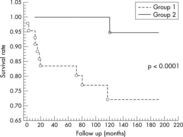
One hundred and fifty one patients were followed up over a mean of 7 (4.3) years. There were 11 fatal events and within the first five years the sudden death-free survival rate was 95% in the overall cohort. The incidence of sudden death was 10.1/1000 person/year and annual mortality was 1%. Comparing group 1 with 2, five year survival was 83% v 100% (fig 1); incidence of sudden death was 31.2 v 1.4/1000 person/year; and mean annual mortality was 3.1% v 0.1% (p < 0.001 for all differences).
Figure 1.
Comparison of sudden death-free survival rate between groups 1 and 2.
The combined echocardiographic criteria (S/LVDd index > 0.5 with reversed septal curvature) predicted sudden death with 91% sensitivity and 76% specificity. The positive and negative predictive accuracy was 23% and 97%, respectively.
An age adjusted univariate and multivariate Cox regression model showed that reversed septal curvature solely (univariate RR 20.2, 95% CI 2.56 to 159, p < 0.004; multivariate RR 11.8, 95% CI 1.15 to 121, p < 0.038) or together with S/LVDd index > 0.5 (univariate RR 33.2, 95% CI 4.21 to 264, p < 0.001; multivariate RR 36.8, 95% CI 3.25 to 417, p < 0.004) was a potent predictor of sudden death. Only in univariate analysis, significant septal hypertrophy was a predictor of sudden death but only if the cut off value was lower than previously reported—that is, 25 mm (RR 7.86, 95% CI 2.04 to 30.3, p < 0.003) instead of 30 mm (RR 0.87, 95% CI 0.10 to 7.13, p = 0.89).
DISCUSSION
Small LV cavity was related to syncope5 and abnormal blood pressure response to exercise. The small LV cavity may facilitate exercise induced hypotension through activation of LV mechanoreceptors. An exaggerated activation of LV mechanoreceptors may induce a withdrawal of sympathetic tone to the peripheral resistance vessels, resulting in sudden hypotension and haemodynamic collapse. A small LV cavity may predispose to hypotensive syncope during LV low input–low output failure triggered by tachycardia.5 It has been suggested that the combination of low filling volume and non-sustained ventricular tachycardia can be particularly detrimental, leading to sudden depression of cardiac output with profound hypotension.5 In these circumstances ventricular fibrillation may develop as the summative effect of myocardial ischaemia (due to hypotension mediated decrease of coronary perfusion pressure) and electrical instability (non-sustained ventricular tachycardia). Also, supraventricular tachycardia predisposes to hypotensive syncope in the presence of a small LV cavity due to the development of similar low input–low output failure.
In a pathomorphological study7 advanced abnormality in myocardial architecture (corresponding to reversed septal curvature) was common in patients who died at a mean age of 25 years. In contrast, most patients who were older than 65 years when they died had a normal circular unit surrounding the LV cavity as a morphological marker of non-reversed septal configuration.
In conclusion, the proposed combination of echocardiographic parameters reflecting a significant morphological imbalance between hypertrophied septum with reversed curvature and narrowed LV cavity allows retrospective identification of a subgroup of patients with HCM who have an increased risk of sudden death. These preliminary results need to be confirmed by prospective analyses.
Abbreviations
HCM, hypertrophic cardiomyopathy
LV, left ventricular
RR, relative risk
S/LVDd, ratio of septal thickness to left ventricular diastolic diameter
REFERENCES
- 1.Elliott PM, Poloniecki J, Dickie S, et al. Sudden death in hypertrophic cardiomyopathy: identification of high risk patients. J Am Coll Cardiol 2000;36:2212–8. [DOI] [PubMed] [Google Scholar]
- 2.McKenna WJ, Behr R. Hypertrophic cardiomyopathy: management, risk stratification and prevention of sudden death. Heart 2002;87:169–76. [DOI] [PMC free article] [PubMed] [Google Scholar]
- 3.Spirito P, Bellone P, Harris K, et al. Magnitude of left ventricular hypertrophy and risk of sudden death in hypertrophic cardiomyopathy. N Engl J Med 2000;342:1778–85. [DOI] [PubMed] [Google Scholar]
- 4.Elliott P, Blanes JR, Mahon N, et al. Relation between severity of left-ventricular hypertrophy and prognosis in patients with hypertrophic cardiomyopathy. Lancet 2001;357:420–4. [DOI] [PubMed] [Google Scholar]
- 5.Nienaber CA, Hiller S, Spielmann RP, et al. Syncope in hypertrophic cardiomyopathy: multivariate analysis of prognostic determinants. J Am Coll Cardiol 1990;15:948–55. [DOI] [PubMed] [Google Scholar]
- 6.Lever HM, Karam RF, Currie PJ, et al. Hypertrophic cardiomyopathy in the elderly: distinctions from the young based on the cardiac shape. Circulation 1989;79:580–9. [DOI] [PubMed] [Google Scholar]
- 7.Kuribayashi T, Roberts WC. Myocardial disarray at junction of ventricular septum and left and right ventricular free walls in hypertrophic cardiomyopathy. Am J Cardiol 1992;70:1333–40. [DOI] [PubMed] [Google Scholar]


