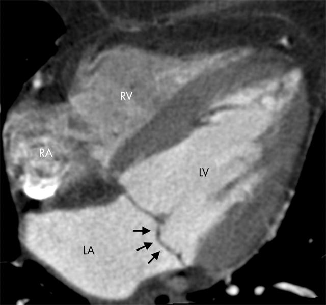
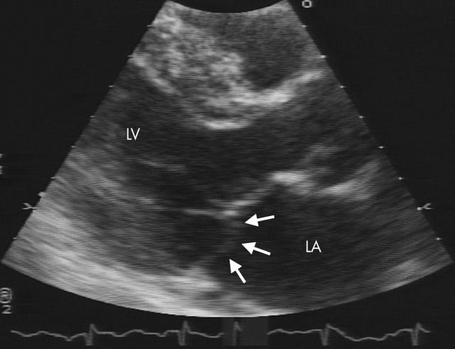
A 67 year old woman who had conflicting findings on stress echocardiography and myocardial scintigraphy with technetium Tc 99m sestamibi presented for participation in a clinical trial of minimally invasive coronary angiography with contrast enhanced, retrospectively gated, multislice (16 slice) spiral computed tomography (MSCT). MSCT images gated in diastole showed severe coronary calcification but no luminal narrowing of > 70% in coronary segments that could be assessed. As per the study protocol, 20 transaxial MSCT images every 5% of the R-to-R interval were reconstructed in horizontal long axis at one intermediate level for qualitative assessment of global and regional left ventricular function. MSCT image reconstructions showed previously unrecognised displacement of the posterior mitral valve leaflet into the left atrium during systole. An echocardiogram, interpreted by a cardiologist unaware of the MSCT findings, subsequently confirmed posterior mitral valve leaflet prolapse. Associated mild mitral regurgitation was documented, and the patient was advised of the need for endocarditis prophylaxis.
For minimally invasive coronary angiography, MSCT data are usually reconstructed in diastole to minimise the influence of cardiac motion on image quality. Using image data reconstructed at other time points in the cardiac cycle can result in common, clinically relevant diagnoses such as abnormalities of mitral valve morphology and function.
Supplementary Material
Figure 1.
Multiplanar reformation of multislice spiral computed tomographic images in horizontal long axis orientation. The reconstruction window begins at 25% of the R-to-R interval, which corresponds to the late phase of isovolumic contraction. Atrial displacement of the posterior mitral valve leaflet (arrows) was most pronounced at this time point. LA, left atrium; LV, left ventricle; RA, right atrium; RV, right ventricle.
Figure 2.
Magnified echocardiographic parasternal long axis view, end systolic frame, shows the posterior mitral valve leaflet prolapse (arrows).
Associated Data
This section collects any data citations, data availability statements, or supplementary materials included in this article.


