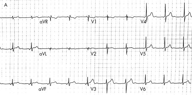
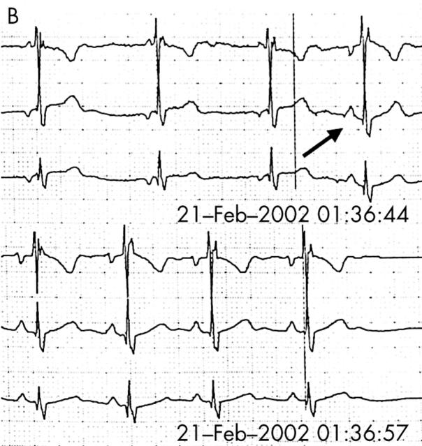
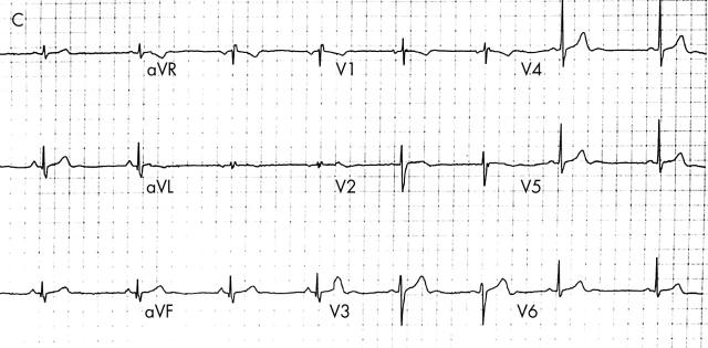
A 28 year old male patient was admitted to the emergency room after a car accident with high velocity impact. Even though the patient did not experience any chest pain, an ECG was obtained and biochemical cardiac markers were determined to exclude cardiac contusion, associated with rapid deceleration trauma. The ECG (panel A) revealed an atrial rhythm with a PR interval of 10 ms; 1 mm ST segment elevation was present in leads II, III, and aVF. Biochemical markers were within normal limits. Because the ECG abnormalities could be caused by transmural ischaemia, echocardiography was performed which did not show any wall motion abnormalities. During observation the atrial rhythm converted spontaneously to sinus rhythm (panel B, leads V1, II, and III are shown), and immediate resolution of the ST segment elevation occurred (panel C).
The immediate resolution indicated a close correlation between the ST segment elevation and the ectopic atrial rhythm. In normal conditions, atrial repolarisation (Ta) is represented by a slow wave with a direction opposite to that of the P wave. Generally Ta is not visible in the ECG, as it is a small electrical event and is often incorporated in the QRS complex. Ectopic atrial activation inferior in the atrium results in a negative P wave, and consequently induces a positive Ta wave. The latter in combination with a short PR interval can modify the ST segment. Although it is a rare phenomenon, it can lead to elevation of the segment.


