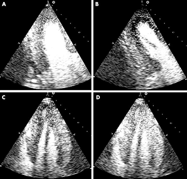
Figure 13.
Contrast destruction-reperfusion study during dobutamine stress echocardiography in a patient with 70% mid left anterior descending coronary artery lesion and a severely diseased right coronary artery with a 90% lesion. Panels A and B are end systolic apical two chamber views at rest (A) three beats post-destruction and at peak stress (B) one beat post-destruction. A significant sub-endocardial defect is seen in the anterior, apical, and inferior walls. Panels C and D are both apical four chamber views at peak stress. Panel C is one beat post-destruction and shows an apical and septal sub-endocardial defect. It also demonstrates aneurysmal pouching of the apex which was even more evident on real time images. Panel D is three beats post-destruction and the sub-endocardial defects have filled in although the apical shape distortion is still evident (post-ischaemic stunning).

