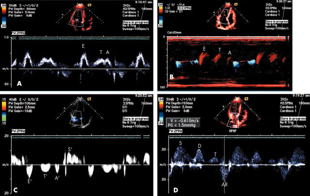
These images show a triphasic pattern of left ventricular (LV) filling in a symptomatic 15 year old patient with non-obstructive hypertrophic cardiomyopathy. This pattern of LV filling is unusual. The sequence clearly demonstrated three separated filling waves. The underlying mechanism seems to be a mixed pattern of restrictive filling and delayed impaired relaxation. The tissue Doppler image in this case is compatible with the recognised diastolic asynchronisation found in hypertrophic cardiomyopathy. The third annulus longitudinal movement may be caused by an extra myocardial relaxation, with suction capacity.
Figure 1.
Panel A: mitral Doppler inflow. Between early (E) and atrial (A) waves, there is a third wave (T) indicative of a mid diastolic filling. Panel B: M colour flow mapping. The mid diastolic filling wave (T) is also evident, and well separated from E and A waves. Panel C: pulsed tissue Doppler of the lateral mitral annulus. During left ventricular systole, the longitudinal movement is toward the apex (S’). In diastole, the early velocity (E’) is low (< 10 cm/s), reflecting impaired relaxation. Before the late atrial velocity (A’), there is a clear and discriminated mid diastolic annular movement (T’). Panel D: pulmonary venous Doppler flow. The systolic (S) and early diastolic (D) waves are well delineated. The atrial reverse wave (AR) has a high peak velocity (> 50 cm/s). Between the two diastolic waves (D and AR), there is a third (T) wave.


