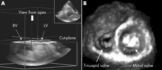
A 56 year old man was admitted for evaluation of exertional dyspnoea. His medical history was significant for rheumatic mitral and aortic valve disease. A transthoracic three dimensional (3D) echocardiography examination was performed using a Philips Sonos 7500 (Philips Medical Systems, Eindhoven, the Netherlands) and a new 4 MHz, 4X matrix transducer capable of providing real time two dimensional (2D) and live 3D transthoracic images. Pyramidal shaped full volume 3D images were acquired with ECG gating by asking the patient to hold his breath for a few seconds. The system permits cropping of 3D images in three different orthogonal axes (lateral, elevational, and depth). The image was cropped using an elevational cutting plane to obtain the profile of the tricuspid valve area viewed from the apex perspective. Three dimensional images were then digitally stored on optical disk and transferred into a dedicated computer (Tom-TEC, Echo-View) for measurements. A valve area of 1.4 cm2 was measured.
Tricuspid stenosis is a rare clinical condition with rheumatic disease accounting for about 90% of all cases. Two dimensional echocardiography permits definitive diagnosis of tricuspid stenosis showing thickening and shortening of the valve leaflets. Nevertheless, unlike evaluation of mitral stenosis, short axis 2D imaging of the valve orifice is rarely feasible. In this patient, an image of tricuspid valve area was easily obtained using 3D transthoracic echocardiography. To our knowledge these images have not been reported previously.
Figure 1.
Using four ECG triggered cardiac beats with the patient breath holding, four subvolumes were time aligned to render a pyramidal shaped full volume image (panel A, top right corner). To obtain the tricuspid valve area from the ventricular perspective, the three dimensional full volume was cropped using the elevational plane from the apex.


