Abstract
Blunt trauma is uncommonly followed by intracardiac valvar injuries. The resulting valvar insufficiency rapidly or progressively leads to congestive heart failure or death unless surgically corrected. Three patients with sustained blunt chest trauma were found to have two aortic valve and one mitral valve ruptures. They had variable clinical courses. However, after the diagnosis was established, surgical intervention was attempted promptly, which consisted of two aortic valve replacements and one mitral valvoplasty. Their postoperative courses were uneventful. Careful observation and repeated physical examination, aided by echocardiography, were required after the blunt chest trauma.
Keywords: aortic valve, mitral valve, blunt trauma
Cardiothoracic blunt trauma following trauma is very common (30%) in civilian life1; however, intracardiac organic injury resulting from blunt chest trauma is rare. When the aortic valve is traumatically injured, it usually has a tear or avulsion on the cusp or on a commissure as well. The injury is often combined with ascending aorta trauma.2,3 The mitral apparatus injury may be caused by papillary muscle rupture or dysfunction, chordae tendineae rupture, or leaflet tears.3 Here we present our surgical experiences in the management of such traumatic valvar lesions after blunt chest injury.
PATIENT 1
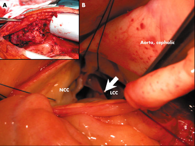
A 65 year old male truck driver, who was involved in a high velocity road accident, sustained a chest contusion and left tibiofibular fractures. On physical examination, the patient was clear with stable vital signs. No cardiac murmur was heard on auscultation. Laboratory data were creatine kinase concentration of 4995 U/l and creatine kinase MB fraction of 12.32 U/l. The ECG showed normal sinus rhythm with a myocardial ischaemia pattern. Transthoracic echocardiography showed adequate left ventricular performance and no pericardial effusion. Two days later, dyspnoea on exertion was noted. On auscultation, his heart beat was irregular and a grade IV/VI diastolic murmur was heard over the aortic area. A cardiac catheter and angiography showed a critical left circumflex coronary occlusion lesion with moderate aortic regurgitation. During the procedures, he had respiratory distress and an endotracheal tube was inserted. He was referred to our hospital for further examination. Repeat transoesophageal echocardiography showed moderate to severe aortic regurgitation. The patient received an urgent operation under the impression of aortic valve injury. When his chest was opened through midline sternotomy, manubrium-sternum fractures and right ventricle ecchymosis were noted (fig 1A). A standard cardiopulmonary bypass was instituted and a transverse aortotomy was done. Cold blood cardioplegia was given through the coronary artery orifices. Avulsion of the left coronary cusp through the mid portion was seen (fig 1B). The aortic cusps were excised and a 23 mm Edwards MIRA valve prosthesis was inserted (Edwards Lifesciences, Irvine, California, USA). He is regularly followed up at outpatient clinics with good recovery.
Figure 1.
Patient 1. (A) Manubrium-sternum fractures were seen after median sternotomy. (B) Intraoperative view of the aortic valve. Avulsion of left coronary cusp (LCC) through the mid-portion (white arrow) was seen. NCC, non-coronary cusp.
PATIENT 2
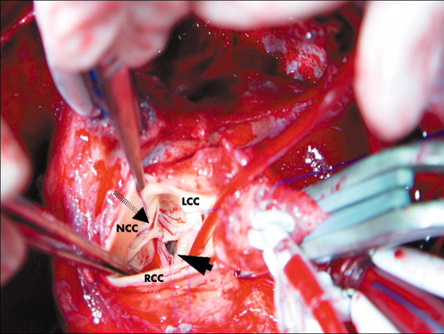
A 50 year old man fell down from a scaffolding about 8 m high. He was clear in consciousness. Physical examination showed tachycardia without murmur, decreased left side breathing sound, left flail chest with haemopneumothorax, and an L3 compression fracture. Endotracheal intubation with ventilator support was instituted and a left side chest tube was inserted immediately. Transthoracic echocardiography showed adequate left ventricular performance and trivial aortic and mitral regurgitation. The chest tube was removed on day 10; however, the left pleural effusion recurred on day 17, so another chest tube was inserted. Unfortunately, the patient bled profusely after the chest tube was inserted. Exploratory thoracotomy was performed immediately. The bleeding was caused by a tiny tear in the right ventricle, which was repaired. On day 20, a remarkable diastolic murmur (grade III/VI) was auscultated over the aortic area. Repeated transthoracic echocardiography showed flail aortic valve with severe aortic regurgitation, confirmed by cardiac catheterisation. Elective surgery was performed on day 32. When the pericardium was opened, extensive adhesions were found. After cardiopulmonary bypass and cardioplegic arrest were established, the aorta was opened transversely. A 2 cm intimal tear with a flap was noted at the aorta above the non-coronary cusp area. An avulsion of the left side of the right coronary cusp was seen about 8 mm along where the annulus was found (fig 2). After the aorta was repaired with a 4-0 prolene continuous suture, the aortic leaflets were excised and replaced by a 23 mm Edwards MIRA valve prosthesis (Edwards Lifesciences). His postoperative course was uneventful and he is regularly followed up at outpatient clinics with good recovery.
Figure 2.
Patient 2. Intraoperative view of the aortic valve. An intimal tear was noted at the aorta above the NCC area (dotted arrow). There is an avulsion of the left side of the right coronary cusp (RCC) about 8 mm along where the annulus (large arrow) was found.
PATIENT 3
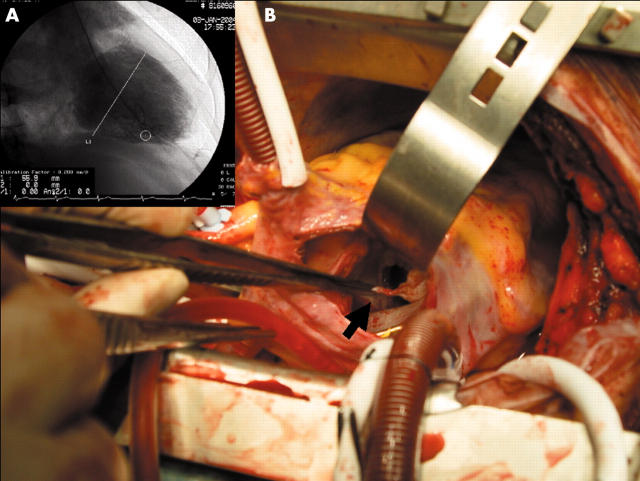
A 64 year old healthy male worker fell from a 2 m height. He noted chest tightness at that time but the patient did not pay attention to it. One month later, he had dyspnoea and a gradually worsening cough with sputum. He visited the regional hospital for help. On auscultation, a significant pansystolic murmur was heard. Echocardiography showed severe mitral regurgitation caused by chordal rupture of the anterior leaflet of the mitral valve, corresponding to the A2–A3 segments (Carpentier’s classification). Under the impression of traumatic mitral regurgitation, he was referred to our hospital for surgical intervention. After the institution of cardiopulmonary bypass and cardioplegic arrest, the left atrium was opened. The primary chordae of the A2–A3 segments were found to be ruptured at the level of the tip of the posterior papillary muscle (fig 3). The papillary muscle looked normal on inspection. A flap of facing normal posterior leaflet with its chordae was removed. It was transferred and sutured to the corresponding segments of the anterior leaflet. In addition, a bilateral commissuroplasty was performed. The patient was weaned off bypass easily. Intraoperative transoesophageal echocardiography showed trivial mitral regurgitation with good leaflet excursion. He is regularly followed up at outpatient clinics. The latest echocardiographic study found only trivial mitral regurgitation.
Figure 3.
Patient 3 (A) Cardiac catheterisation shows severe mitral regurgitation on left ventriculography. (B) Intraoperative view of the mitral valve through the septum. Primary chordae of the A2–A3 segments were ruptured at the level of the tip of the posterior papillary muscle (arrow).
DISCUSSION
Blunt chest trauma occurs most often after high speed motor vehicle crashes or after falls.3,4 Myocardial contusion, a common injury that may impair ventricular contraction and lead to arrhythmia, occurs in up to 16–76% of patients after blunt chest trauma.5 Valve injury is rare; only a few cases have been reported in the literature, with the aortic valve most involved, followed by the mitral and tricuspid valves. The mechanism that accounts for both mitral and aortic injuries is a sudden increase in intracardiac pressure during a vulnerable phase (especially during isovolumetric contraction) of the cardiac cycle.6 For the aortic valve, it is the early diastole, when a tremendous pressure gradient can be generalised across a competent aortic valve.7 The mitral valve and subvalvar apparatuses are vulnerable during late diastole and early systole, when the delivered impact force suddenly changes the full flooded ventricle and stretches the mitral apparatus.4 A fractured sternum, as seen in our patients, is a common feature in these scenarios. Tear or avulsion from the annulus of one aortic valve cusp, especially in the non-coronary cusp, is the most frequently observed aortic valve lesion3; it is different from that in our patients. The most common mitral lesion is rupture of the papillary muscles, followed by the chordae tendineae and a leaflet tear,8 as in our case.
A diagnosis of traumatic valvar injury often is suggested by acute or progressive heart failure or a new heart murmur and history of blunt chest trauma.7 Clinical findings of patients vary widely, from asymptomatic to acute cardiogenic shock. Symptoms are often acute in patients with aortic injury, as in our patient 1. These symptoms can also be present subacutely, as with patient 2. Mitral valve injury usually has an asymptomatic stage initially, as in our patient 3. It may be partial papillary muscle or chordae damage at first but under subsequent wall stress, these injured apparatuses lead to complete disruption and aggravated symptoms.3 Echocardiography is the non-invasive test of choice to diagnose these conditions.9
The indications for aortic valve replacement versus aortic valve repair depend on the extent of the lesion in the damaged cusp or the number of cusps involved. Although successful repair of these injuries has been reported, there is still a long term outcome that is not warrantable.4 Aortic valve replacement seemed the best choice for these patients. For the mitral valve, surgical strategies, including a variety of repairs and replacement, are justified by the extent and location of damage, the interval from injury to operation, accurate analysis of the mitral valve and its subvalvar apparatus, and the surgeon’s expertise. Ruptured chordae have been directly sutured to the free ventricular wall, replaced with an autologous fascia lata graft or artificial material, or repaired by the chordae transfer technique, as in our case.
In conclusion, aortic and mitral lesions resulting from blunt chest trauma are almost exceptional. Patients with recent or a history of high energy blunt chest trauma, including traffic accidents and falling from heights, must be promptly and repeatedly examined physically and by echocardiography to rule out either isolated or combined intracardiac lesions.
REFERENCES
- 1.Symbas PJ, Horsley WS, Symbas PN. Rupture of the ascending aorta caused by blunt trauma. Ann Thorac Surg 1998;66:113–7. [DOI] [PubMed] [Google Scholar]
- 2.West O, Vanderbush E, Anagnostopoulos CE. Traumatic rupture of the aortic valve and ascending aorta diagnosed by transesophageal echocardiography. J Cardiovasc Surg (Torino) 1999;40:671–3. [PubMed] [Google Scholar]
- 3.Bernabeu E, Mestres CA, Loma-Osorio P, et al. Acute aortic and mitral valve regurgitation following blunt chest trauma. Int Cardiovasc Thorac Surg 2004;3:198–200. [DOI] [PubMed] [Google Scholar]
- 4.Halstead J, Hosseinpour AR, Wells FC. Conservative surgical treatment of valvular injury after blunt chest trauma. Ann Thorac Surg 2000;69:766–8. [DOI] [PubMed] [Google Scholar]
- 5.Pretre R, Chilcott M. Blunt trauma to the heart and great vessels. N Engl J Med 1997;336:626–32. [DOI] [PubMed] [Google Scholar]
- 6.Banning AP, Pillai R. Non-penetrating cardiac and aortic trauma. Heart 1997;78:226–9. [DOI] [PMC free article] [PubMed] [Google Scholar]
- 7.Egoh Y, Okoshi T, Anbe J, et al. Surgical treatment of traumatic rupture of the normal aortic valve. Eur J Cardiothorac Surg 1997;11:1180–2. [DOI] [PubMed] [Google Scholar]
- 8.Reardon MJ, Conklin LD, Letsou GV, et al. Mitral valve injury from blunt trauma. J Heart Valve Dis 1998;7:467–70. [PubMed] [Google Scholar]
- 9.Zakynthinos EG, Vassilakopoulos T, Routsi C, et al. Early- and late-onset atrioventricular valve rupture after blunt chest trauma: the usefulness of transesophageal echocardiography. J Trauma 2002;52:990–6. [DOI] [PubMed] [Google Scholar]


