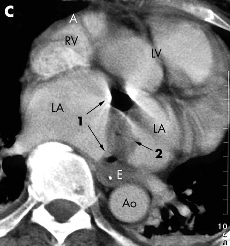
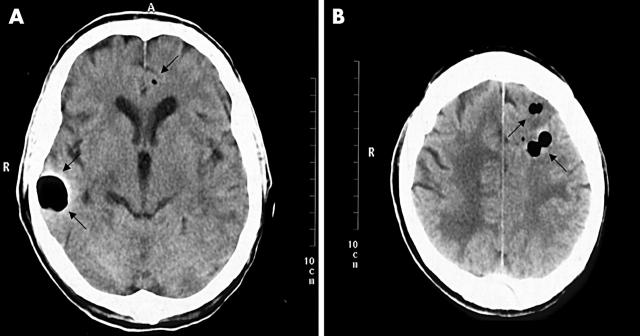
A 66 year old white man underwent mitral valve replacement in another city for severe mitral regurgitation. Surgery included a Maze procedure due to persistent atrial fibrillation. The postoperative course was uncomplicated. Two weeks after discharge the patient collapsed. Being unconscious he had to be intubated. An initial computed tomographic (CT) scan and magnetic resonance imaging of the head was normal. A transoesophageal echocardiogram showed good function of the prosthetic valve. The patient developed severe sepsis. In a control CT scan two weeks later multiple intracerebral air emboli as well as multiple cerebral infarctions were observed (panels A and B). A CT scan of the thorax and the abdomen revealed a thrombus and free air in the left atrium (panel C) as well as embolic lesions in both kidneys and the spleen. Finally, an oesophagoatrial fistula was diagnosed by oesophagoscopy and palliative stenting was performed. The patient died in septic shock with the neurological picture of an acinetic mutism. To our knowledge this is the first case reporting multiple air emboli caused by an oesophagoatrial fistula after cardiac surgery involving the Maze procedure. In case of postoperative sepsis and neurological deficits one should think of this form of complication.
Figure 1.
Cerebral computed tomographic (CT) scan showing multiple air emboli (arrows).
Figure 2.

Thoracic CT scan revealing free air (1) and a large thrombus (2) in the left atrium. Ao, aorta; E, oesophagus; LA, left atrium; LV, left ventricle; RV, right ventricle.


