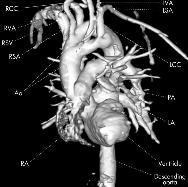
We present a case of a 73 year old woman with double aortic arch and separate origin of the right common carotid and right subclavian arteries, found coincidentally during cardiac angiography. She has a history of coronary artery disease and atherosclerotic renal artery occlusion. Medical history includes indigestion (relieved by proton pump inhibition), deep vein thrombosis, pulmonary embolic disease, coronary bypass grafting, and a minor stroke. The image (three dimensional reconstructed magnetic resonance contrast angiography) demonstrates the double aortic arch. The right common carotid and right subclavian arteries arise separately from the posterior arch and the left common carotid and left subclavian from the anterior arch. The right and left vertebral arteries arise from the corresponding subclavian arteries. The right vertebral artery is stenotic and shows post-stenotic dilatation. Both aortic lumina are similar in calibre and intact. There was no dysphagia or stridor and there was no tracheal compression identified on native images. This observation suggests that the incidence and prevalence of this rare condition may be underestimated.
Figure 1.
Ao, aorta; LCC, left common carotid; LSA, left subclavian artery; LVA, left vertebral artery; RCC, right common carotid; RSA, right subclavian artery; RVA, right vertebral artery.


