A 40 year old Asian male collapsed while attending a wedding in India. He initially assumed that his drinks had been “spiked”. He attended a hospital in India when further episodes of dizziness occurred. An ECG showed a brief run of a broad complex tachycardia. Haematology and biochemistry were normal as was the chest radiograph and a transthoracic echocardiogram. On returning to the UK he was found to have self terminating runs of polymorphic ventricular tachycardia at rates of 190 beats/min. A 12 lead ECG was normal as was a repeat echocardiogram. Coronary angiography showed no evidence of obstructive coronary artery disease.
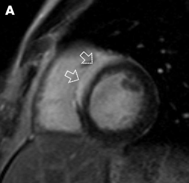
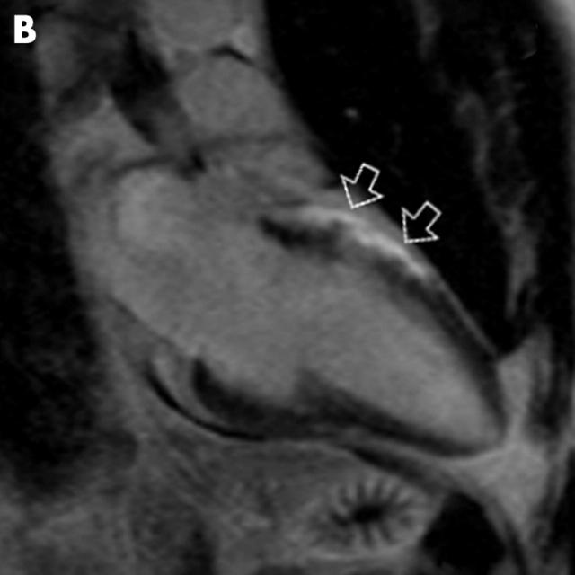
A magnetic resonance imaging (MRI) scan was performed with a view to excluding a structural abnormality and specifically right ventricular dysplasia. Both ventricles were of normal size and function. However, on the delayed contrast enhanced images, there was hyperenhancement of the anteroseptal segments of the basal and mid thirds of the left ventricle in an epicardial distribution (panels A and B). Enlarged lymph nodes were seen in a pre-aortic location on the T1 weighted images. Based on these features a provisional diagnosis of sarcoidosis was suggested. Open biopsy of a submandibular lymph node, located on ultrasound, confirmed the presence of sarcoidosis. No pulmonary manifestations of sarcoidosis were seen on the computed tomographic scan.
An automated implantable cardioverter-defibrillator was implanted. The patient remains well on sotalol, 360 mg/day, and oral prednisolone, 20 mg/day.
Cardiac MRI should be considered in patients with rhythm disturbance when the echocardiogram appears to show an entirely normal heart.


