Abstract
A 53 year old woman presented with a lump in the inner lower quadrant of the left breast. Histological examination of the breast tumour confirmed that the lesion was a mammary hamartoma. Carcinoma with foci of microinvasion was observed in the lobules of the hamartoma concomitant with the intraductal spread of lobular carcinoma. Immunohistochemically, the cancer cells were negative for β-catenin, which generally stained normal breast ducts and ductal carcinomas. This is only the sixth case of breast carcinoma arising in a mammary hamartoma to be reported and, moreover, the fourth case of lobular carcinoma occurring within a hamartoma. Despite the apparent rarity of this case, pathologists should be aware of the possibility of carcinomas arising within mammary hamartomas.
Keywords: lobular carcinoma; intraductal spread; hamartoma, breast
CASE REPORT
A 53 year old Japanese woman discovered a lump in the lower inner quadrant of her left breast and visited the first department of surgery, Kochi Medical School Hospital in July 2000. A physical examination revealed a palpable soft mass measuring 6 × 5 cm, but no axillary lymphadenopathy. Ultrasonography revealed an iso-echoic solid mass with a capsule suspected to be a lipomatous tumour. Mammography revealed a circumscribed mass with a thin pseudocapsule, but neither spiculation nor microcalcification, which are often seen in typical breast carcinomas. The patient underwent a tumorectomy in the same month. The next month, she also underwent a partial mastectomy on the suspicion of remaining carcinoma in the surgical margin. However, no residual tumours were found on histological examination after the operation. Her convalescence was uneventful four months after surgery.
PATHOLOGY
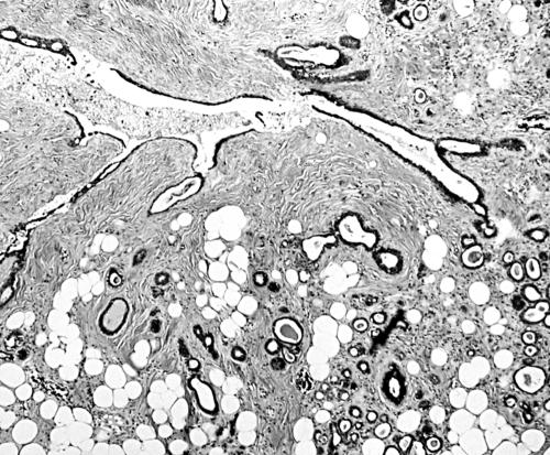
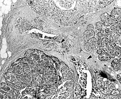
The tumour located in the lower inner quadrant of the left breast was a well circumscribed mass of soft consistency. Macroscopically, the cut surface of the 6 × 5 × 3 cm tumour was yellowish. In addition, hard nodules measuring 0.3 × 0.3 cm in diameter were identified on the serial cut surface. Histological examination confirmed that the lesion was a mammary hamartoma consisting of epithelial components, adipose tissue, and dense connective tissue. Unlike fibroadenoma, both ducts and lobules were present (fig 1). Moreover, a portion of the lobular epithelium showed malignant transformation with foci of microinvasion. Carcinomatous changes were also seen in the ducts (fig 2). The cancer cells generally lacked cohesiveness and no tubular formation was identified. Immunohistochemically, all the tumour cells from our case and two typical lobular carcinomas acting as negative controls for β-catenin immunostaining were completely negative for β-catenin (clone E-5; 1/50 dilution; Santa Cruz Biotechnology, Santa Cruz, California, USA), whereas normal breast ducts and three other invasive ductal carcinomas acting as positive controls showed strong membranous positivity for β-catenin. These findings confirmed that the carcinoma occurring in the mammary hamartoma was lobular in nature.
Figure 1.
Microscopic findings of the background lesion (haematoxylin and eosin stain). A random mixture of epithelial and mesenchymal components can be seen. Periductal fibrosis is evident.
Figure 2.
Microscopic examination of nodules reveals the proliferation of tumour cells in the lobules, indicative of lobular carcinoma (haematoxylin and eosin stain). Microinvasion (arrow) and intraductal spread (arrowhead) are also apparent.
“A portion of the lobular epithelium showed malignant transformation with foci of microinvasion”
DISCUSSION
Mammary hamartoma was first identified by Arrigoni et al in 1971.1 This tumour is generally asymptomatic, with patients complaining of a lump in the symptomatic cases.2–4 The breast hamartoma is a tumour that may be underdiagnosed by pathologists because it has all the constituents of normal breast tissue.3,4 Macroscopically, it is well circumscribed.1–4 Light microscopy shows a varied combination of epithelial and stromal elements and it is easily differentiated from fibroadenoma by the presence of both lobular structures and ducts.1–4 Among mammary hamartomas previously reported in the literature, only five cases in which the hamartoma contained carcinoma have been described. Among these, two cases were lobular carcinomas and two cases were ductal carcinomas.5–7 Recently, Mester et al reported a case with both ductal and lobular carcinomas.8 The age (mean, 45 years) of patients with hamartoma with carcinoma is generally lower than that (mean, 60 years) of patients with usual hamartomas.2,5–8
In cases where it is difficult to distinguish whether the breast carcinoma is ductal or lobular, immunohistochemical tests using antibodies against β-catenin or E-cadherin can be useful.9 The proliferation of carcinoma mainly in the lobules and the negative reaction of neoplastic cells for β-catenin as seen in our present case confirms a diagnosis of lobular carcinoma.
In conclusion, we report here a rare case of lobular carcinoma showing intraductal spread arising in a mammary hamartoma. Despite the apparent rarity of such a lesion, pathologists should be aware of the possibility of carcinomas arising within mammary hamartomas. Furthermore, we would like to emphasise the importance of careful macroscopic and microscopic examination of breast tumours to enable the presence of extremely small carcinomas to be determined, even in the more unusual cases such as mammary hamartomas.
Take home messages.
Although rare, carcinomas can arise within mammary hamartomas
Careful macroscopic and microscopic examination of breast tumours should be undertaken to enable the presence of very small carcinomas to be determined
Acknowledgments
We gratefully ackowledge the excellent technical assistance provided by Mr T Tokaji and Ms E Miyazaki at the first department of pathology, Kochi Medical School. We are also grateful to Mr D Blake for correcting the English in our manuscript.
REFERENCES
- 1.Arrigoni MG, Dockerty MB, Judd ES. The identification and treatment of mammary hamartoma. Surg Gynecol Obstet 1971;133:577–82. [PubMed] [Google Scholar]
- 2.Daya D, Trus T, D'souza TJ, et al. Hamartoma of the breast, an underrecognized breast lesion. A clinicopathologic and radiographic study of 25 cases. Am J Clin Pathol 1995;103:685–9. [DOI] [PubMed] [Google Scholar]
- 3.Fisher CJ, Hanby AM, Robinson L, et al. Mammary hamartoma—a review of 35 cases. Histopathology 1992;20:99–106. [DOI] [PubMed] [Google Scholar]
- 4.Linell F, Ostberg G, Soderstrom J, et al. Breast hamartoma. An important entity in mammary pathology. Virchows Arch A Pathol Anat Histol 1979;383:253–64. [DOI] [PubMed] [Google Scholar]
- 5.Toft M, Al-Harris JA. Lobular carcinoma in situ occurring in adenolipoma of the breast. Report of a case. Acta Radiol 1982;23:503–5. [DOI] [PubMed] [Google Scholar]
- 6.Coyne J, Hobbs FM, Boggis C, et al. Lobular carcinoma in a mammary hamartoma. J Clin Pathol 1992;45:936–7. [DOI] [PMC free article] [PubMed] [Google Scholar]
- 7.Anani PA, Hessler Ch. Breast hamartoma with invasive ductal carcinoma. Report of two cases and review of the literature. Pathol Res Pract 1996;192:1187–94. [DOI] [PubMed] [Google Scholar]
- 8.Mester J, Simmons RM, Vazquez MF, et al. In situ and infiltrating ductal carcinoma arising in a breast hamartoma. Am J Roentgenol 2000;175:64–6. [DOI] [PubMed] [Google Scholar]
- 9.De Leeuw WJF, Berx G, Vos CBJ, et al. Simultaneous loss of E-cadherin and catenins in invasive lobular breast cancer and lobular carcinoma in situ. J Pathol 1997;183:404–11. [DOI] [PubMed] [Google Scholar]


