Abstract
An 8 year old girl with cystic fibrosis presented with a pulmonary exacerbation from which Burkholderia cepacia was cultured. Subsequent polymerase chain reaction restriction fragment length polymorphism analysis of the recA gene suggested the presence of B cepacia Genomovar V (Burkholderia vietnamiensis); however, on subsequent sequence typing, this isolate was confirmed as B cepacia Genomovar IIIb. This report outlines the potential difficulties in the correct characterisation of the various genomovars within the B cepacia complex of organisms, which has particularly important implications for patient segregation and infection control.
Keywords: Burkholderia cepacia; cystic fibrosis, Genomovar, infection control, recA
Burkholderia cepacia, a Gram negative bacillus, is a saprophyte in soil and river sediments and can cause “slippery skin” or “sour skin” rot in plants, such as onion and garlic. Originally named Pseudomonas cepacia, it was renamed Burkholderia cepacia in 1992, when taxonomists showed that it was sufficiently different to the Pseudomonas species, based on DNA–DNA hybridisation studies and 16S rRNA sequence alignments. Over the past two decades its role as an important human pathogen has emerged and it has been found to have particularly serious consequences for patients with cystic fibrosis (CF), which may result in accelerated pulmonary deterioration, with fatal necrotising pneumonia and bacteraemia.1
Recently, several genomic species or “genomovars” of this species have been described.2 These genomovars are phenotypically indistinguishable, but have sufficient differences in both 16S rRNA phylogeny and DNA–DNA hybridisation to classify them as a separate species. Furthermore, early studies have indicated that there are differences in the virulence of these genomovars, whereby Genomovar III appears to be more virulent than other genomovars.3 To help with their identification, various polymerase chain reaction restriction fragment length polymorphism (PCR-RFLP) systems based on 16S rRNA and the recA4 gene have been described, based on visible differences in DNA banding patterns on agarose gels. Previous workers have reported the potential problems of misidentification using a PCR-RFLP system for Aeromonas spp.5; however, such problems have not been described for the identification of B cepacia, using such a system.
“Early studies have indicated that there are differences in the virulence of these genomovars, whereby Genomovar III appears to be more virulent than other genomovars”
We report a case of potential misidentification of genomovar status in an 8 year old girl with CF which posed a diagnostic and hence an infection control problem.
CASE REPORT
An 8 year old girl (weight, 27.1 kg; height, 128.9 cm) with CF, which was identified at birth by screening and CF genotype (δF508/not identified), presented in November 2000 with a pulmonary exacerbation requiring hospital admission and intravenous antibiotics. She had a history of chronic chest infection with non-mucoid and mucoid Pseudomonas aeruginosa from 3 years and 5 years of age, respectively. Until this admission, she had no previous history of colonisation or infection with B cepacia. On admission, she complained of headaches, poor appetite, cough, and feeling flushed. In addition, she noted a pronounced increase in her sputum production, which was mucopurulent. She had a blood oxygen saturation of 89–90% and a fever. Her lung function tests demonstrated a forced vital capacity of 1.49 litre (85% predicted) and a forced expiratory volume in one second of 1.27 litre (77% predicted). Burkholderia cepacia (API 20NE profile 0467573; identification 99.9% B cepacia) was subsequently cultured from sputum on selective media, at a cell density of 4.375 × 10⁴ colony forming units/g sputum, in addition to a mucoid strain of P aeruginosa. The B cepacia isolate was sensitive to temocillin, ceftazidime, azlocillin, tazocin, imipenem, meropenem, and ciprofloxacin and resistant to gentamicin, tobramycin, and colistin. Molecular characterisation of the B cepacia isolate was requested to help determine how this patient should be optimally segregated on an inpatient basis. Following DNA extraction, the genomic DNA from the isolate was amplified using primers targeting the recA locus. Genomovar status was determined by (1) comparison of RFLP profiles against published profiles generated from reference strains and (2) sequence analysis of the recA amplicon against the GenBank reference database. The RFLP profile suggested the presence of B cepacia Genomovar V (B vietnamiensis) (fig 1); however, on sequence typing and BLAST alignment, this isolate was confirmed as B cepacia Genomovar IIIb. Subsequent management of the patient was appropriate for B cepacia Genomovar IIIb status, whereby the patient was segregated from all other B cepacia colonised patients in the B cepacia inpatient CF unit.
Figure 1.
recA polymerase chain reaction restriction fragment length polymorphism patterns obtained with the query Burkholderia cepacia isolate (lane 1) isolated from an 8 year old girl with cystic fibrosis and B cepacia Genomovar IIIb reference strain (lane 2). Lane 3 contains a negative control (molecular grade water) and lane M contains a molecular weight marker (100 bp; Gibco Life Technologies, Paisley, UK). A and B denote the presence of bands of approximately 175 bp and 100 bp, respectively, in the query isolate.
DISCUSSION
Identification of the correct genomovar status for patients with CF who are infected with B cepacia is essential in terms of managing subsequent infection control. Our case provides an example of common problems encountered in deciding on the genomovar status of this organism in patients with CF. In our case, B cepacia Genomovar V was initially assigned to the query isolate based on comparison of the RFLP patterns against published reference standards (fig 1) because the query isolate had bands around 200 and 100 bp, like B cepacia Genomovar V. In addition, the isolate did not have a band at 150 bp, which is indicative of B cepacia Genomovar III.
Overall, it is important to recognise the consequences for the patient of the misinterpretation of RFLP profiles, for a combination of reasons, as stated above. Minor banding differences can result in changes from Genomovar V to IIIb status, as was illustrated in this case report, with important implications for infection control. Some microbiologists at CF centres (for example, Dublin) believe that it is important to segregate patients with Genomovar IIIs and Genomovar IIs from each other and from other patients infected with B cepacia, but that it is acceptable to manage the other genomovars together (I, IV, and V). In such circumstances, based on the RFLP result (Genomovar V) our patient would have been mixing with other patients with less virulent strains, and thus compromising them in terms of increasing their probability of acquiring the Genomovar IIIb strain, which was the true nature of the isolate.
“We have noted that the interpretation of RFLP banding patterns is subjective and can thus lead to the isolate being classified as the wrong genomovar”
Previously, we have used a PCR-RFLP system in conjunction with the recA gene to determine directly from sputum6 the genomovar status of patients with CF who are colonised with B cepacia. We wish to report several potential problems in the interpretation of RFLP patterns as demonstrated in the above case of the recA PCR system, and suggest practical methods for workers to be able to report results with confidence. We have noted that the interpretation of RFLP banding patterns is subjective and can thus lead to the isolate being classified as the wrong genomovar. This is mainly the result of (1) poor quality genomic DNA for PCR amplification, (2) non-optimisation of the PCR, and (3) insensitive image capture facilities. Each of the genomovars may have two or more RFLP patterns with most of bands—for example, being concentrated in the 300–500 bp range in the case of the HaeIII digestion pattern. In addition, not all bands are of equal intensity, so that weaker bands may be missed, leading to the identification of the wrong genomovar type. It may be argued that the use of well characterised reference strains would eliminate such errors and inaccuracies. Adoption of the former may indeed improve reproducibility; however, it is very difficult to know which reference strains to use as controls, because numerous known RFLP patterns have previously been described for each genomovar type, with the possibility of several unknown profiles existing as a result of the high diversity within this locus. Consequently, many workers may attempt to “best match” RFLP profiles from query isolates with the closest match from reference strains, resulting in potential misidentification of genomovar status.
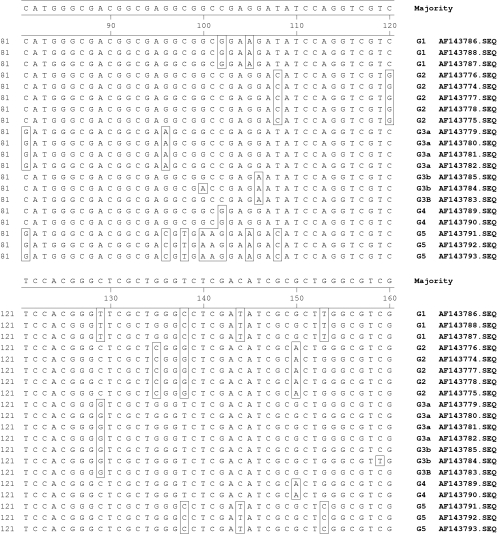
Overall, we would discourage the use of RFLP analysis as the sole system for the differentiation of genomovars and would recommend a sequence typing approach, whereby at least the first 400 bases of the recA gene are characterised and matched against alignment patterns, which have not yet been published (see fig 2 for aligned region position 81–161). Where primary diagnostic laboratories do not have DNA sequencing facilities or access to such facilities, the differentiation of genomovar types may be more reliably determined by carrying out the initial RFLP analysis using stringent controls, including (1) high quality genomic DNA extraction, with a commercially available DNA extraction kit, such as the Roche High Purity PCR kit; (2) empirically optimised PCR conditions; (3) sensitive image capture, using a CCDC camera with an integration facility; (4) the use of DNA molecular weight markers for the range 100–600 bp; (5) electrophoresis of known internal standards and reference genomovar strains; (6) the use of standard precast polyacrylamide matrices, such as ExelGel or CleanGel (Pharmacia Ltd, Amersham, UK); (7) the use of a qualitative gel comparative software system, such as GelCompare or BioNumerics; and (8) confirmation of atypical RFLP profiles by a reference laboratory. In addition, we would recommend confirmation of the genomovar type by species specific PCR, as described previously.4
Figure 2.
Alignments of recA gene sequences from position 81 to 161 of type strains of Genomovars I to V of the Burkholderia cepacia complex demonstrating sequence heterogeneity useful in genomovar identification. G, Genomovar; AF, EMBL accession number.
In conclusion, the generation of reliable results is important in directing appropriate infection control strategies to help control the transmission of this organism to those patients with CF who are not colonised and also to prevent the spread of this organism between patients who are already colonised, because differences in virulence between genomovar types may be important prognostic markers.
Take home messages.
In this patient with cystic fibrosis (CF), restriction fragment length polymorphism (RFLP) mistyped the B cepacia isolate as Genomovar V
Reliable typing is essential to direct appropriate infection control strategies, particularly in CF
RFLP analysis should not be used as the sole system for the differentiation of genomovars and should be supplemented by a sequence typing approach
For those primary diagnostic laboratories that do not have DNA sequencing facilities or access to such facilities, the differentiation of genomovar types may be more reliably determined by carrying out the initial RFLP analysis using stringent controls
Abbreviations
CF, cystic fibrosis
PCR, polymerase chain reaction
RFLP, restriction fragment length polymorphism
REFERENCES
- 1.Govan JR, Hughes JE, Vandamme P. Burkholderia cepacia: medical, taxonomic and ecological issues. J Med Microbiol 1996;45:395–407. [DOI] [PubMed] [Google Scholar]
- 2.Vandamme P, Holmes B, Vancanneyt M, et al. Occurrence of multiple genomovars of Burkholderia cepacia in cystic fibrosis patients and proposal of Burkholderia multivorans sp. nov. Int J Syst Bacteriol 1997;47:1188–200. [DOI] [PubMed] [Google Scholar]
- 3.De Soyza A, Corris PA, Archer L, et al. Pulmonary transplantation for CF; the effect of B. cepacia genomovars on outcomes. Thorax 2000;55(suppl 3):S35. [Google Scholar]
- 4.Mahenenthiralingam E, Bischof, J, Byrne SK, et al. DNA-based diagnostic approaches for identification of Burkholderia cepacia complex, Burkholderia vietnamiensis, Burkholderia multivorans, Burkholderia stabilis, and Burkholderia cepacia Genomovars I and III. J Clin Microbiol 2000;38:3165–73. [DOI] [PMC free article] [PubMed] [Google Scholar]
- 5.Graf J. Diverse restriction fragment length polymorphism patterns of the PCR-amplified 16S rRNA genes in Aeromonas veronii strains and possible misidentification of Aeromonas species. J Clin Microbiol 1999;37:3194–7. [DOI] [PMC free article] [PubMed] [Google Scholar]
- 6.McDowell A, Dunbar K, Moore JE, et al. Speciation of the B. cepacia complex directly from CF sputum. Pediatr Pulmonol 1999;S19:271–2. [Google Scholar]


