Abstract
A 74 year old man had a right hemicolectomy for a caecal adenocarcinoma. Microscopic examination also revealed non-caseating epithelioid granulomatas within the adjacent stroma and some of these granulomata contained centrally located malignant cells. Granulomatous inflammation was not present within the lymph nodes and the importance of this rare occurrence is discussed.
Keywords: caecal adenocarcinoma, epithelioid granulomata
Granulomatous reactions occurring within lymph nodes draining carcinomas are a well known but uncommon occurrence.1 More rarely, granulomas may occur within the stroma of malignancies, a variety of which have been reported including breast, renal, and hepatocellular carcinomas.2–4 However, the occurrence of granulomas within the stroma of colonic carcinomas appears to be much more rare, with only a single case reported in the Japanese literature.5 This case is, to the best of my knowledge, the first case reported in the English literature.
CASE HISTORY AND PATHOLOGY
A 74 year old man was assessed for rectal bleeding, constipation, and weight loss. Six years previously he had a 5 cm moderately dysplastic tubulovillous adenoma removed from the rectum. Colonoscopy revealed three sessile polyps each measuring 2 mm maximum dimension, which were excised from the rectum, proximal sigmoid, and hepatic flexure. In the caecum there was a 45 mm ulcerating tumour encircling the ileocaecal valve with an adjacent 3 mm pedunculated polyp. A 12 mm sessile polyp was also present. A right hemicolectomy was subsequently performed and the patient made an uneventful recovery. Six months later the patient is alive, well, and regaining weight. An abdominal ultrasound showed a normal liver with no focal abnormality. There was no clinical indication of inflammatory bowel disease.
The specimen consisted of caecum and right colon measuring 18 cm in length with a 4 cm piece of terminal ileum. At the ileocaecal valve there was an ulcerated tumour measuring 3.3 × 2.0 cm, which extended through the wall into the adjacent fat.
“The granulomatous reaction may be a response to necrotic material (that is, a foreign body inflammatory response) or an immunological reaction”
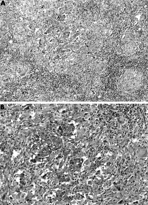
Microscopic examination showed an infiltrating poorly differentiated colonic carcinoma extending into the serosal fat. Focally, there was a granulomatous reaction within the stroma of and adjacent to the carcinoma, and some of the granulomas contained centrally located malignant cells (fig 1). One of 10 lymph nodes contained metastatic mucinous carcinomas, although mucinous elements were not identified in the primary carcinoma. Elsewhere there were several tubulovillous adenomas showing moderate dysplasia. Ziehl-Neelson stains were negative.
Figure 1.
(A) Stromal granulomatous response adjacent to the carcinoma. (B) Granuloma containing centrally located malignant cells.
DISCUSSION
The occurrence of non-caseating granulomas occurring in lymph nodes draining malignant neoplasms is a well documented phenomenon, with cervical and breast carcinomas thought to be the most likely to elicit this response.1,2 Less commonly, non-caseating epithelioid granulomas and metastatic malignancy occur simultaneously within lymph nodes, but this distinctly unusual phenomenon has been described only with metastatic nasopharyngeal carcinoma, seminoma, and malignant melanoma.6,7 An equally rare phenomenon is that of a granulomatous response occurring within the stroma of a variety of carcinomas, including breast, renal, and hepatocellular carcinomas.2–4 A single previous case of sigmoid colon cancer with a sarcoid stromal reaction was described in the Japanese literature.5 However, despite the rarity of these last cases, granulomatous inflammation has been described in association with microinvasive breast carcinomas (predominantly in situ carcinomas with invasive foci measuring < 1 mm) and in relation to microscopic foci of colonic carcinoma within perivascular mesenteric fat.8,9 The granulomatous reaction may be a response to necrotic material (that is, a foreign body inflammatory response) or an immunological reaction, with the granulomatous response within draining lymph nodes representing a response to soluble tumour related antigens and the stromal reaction representing a T cell mediated immunological response to cell surface antigens. Our patient, in whom Crohn's disease and tuberculosis were clinically and immunohistochemically excluded, further supports the contention that this is a very early immunological response to surface antigens.
Take home messages.
The occurrence of granulomas within the stroma of colonic carcinomas is extremely rare and this is the first case reported in the English literature
The granulomatous response within draining lymph nodes may be a response to soluble tumour related antigens and the stromal reaction may be a T cell mediated immunological response to cell surface antigens
Acknowledgments
Thanks to Mrs S Thompson for excellent secretarial assistance and Dr A Curry for the photomicrographs.
REFERENCES
- 1.Gregori HB, Othersen HB, Moore MP. The significance of sarcoid-like lesions in association with malignant neoplasms. Am J Surg 1962;104:577–86. [DOI] [PubMed] [Google Scholar]
- 2.Oberman H. Invasive carcinoma of the breast with granulomatous response. Am J Clin Pathol 1987;88:719–21. [DOI] [PubMed] [Google Scholar]
- 3.Watterson J. Epithelioid granulomas associated with hepatocellular carcinoma. Arch Pathol Lab Med 1982;106:538–9. [PubMed] [Google Scholar]
- 4.Campbell F, Douglas-Jones A. Sarcoid-like granulomas in primary renal cell carcinoma. Sarcoidosis 1993;10:128–31. [PubMed] [Google Scholar]
- 5.Hirano T, Furukawa H, Kawakami Y, et al. A case of sigmoid colon cancer with sarcoid reaction [in Japanese]. Surgical Therapy 1994;71:477–9. [Google Scholar]
- 6.Blackshaw A. Metastatic tumour in lymph nodes. In: Stansfield AG, ed. Lymph node biopsy interpretation. London: Churchill-Livingstone, 1987:380–97.
- 7.Coyne JD, Banerjee SS, Menasce LP, et al. Granulomatous lymphadenitis associated with metastatic malignant melanoma. Histopathology 1996;28:470–2. [DOI] [PubMed] [Google Scholar]
- 8.Coyne JD, Haboubi NY. Micro-invasive breast carcinoma with granulomatous stromal response. Histopathology 1992;20:184–5. [DOI] [PubMed] [Google Scholar]
- 9.Coyne JD, Haboubi NY. Invasive carcinoma with a granulomatous stromal response. Histopathology 1994;25:397–400. [DOI] [PubMed] [Google Scholar]


