Abstract
This report describes a case of angiotropic variant of diffuse large B cell lymphoma within a benign oncocytoma of the lacrimal sac. The occurrence of this rare lymphoma within a benign neoplasm has not been documented previously. An 87 year old woman presented with a swelling over the area of the left lacrimal sac, which histological examination revealed to be an oncocytoma. Many small blood vessels within the tumour were filled with large cytologically atypical cells, which stained positively for leucocyte common antigen and a B cell antigen, CD20, confirming the presence of a large B cell non-Hodgkin’s lymphoma of angiotropic type. Angiotropic lymphoma is a very rare and usually highly aggressive variant of non-Hodgkin’s lymphoma, which classically involves the central nervous system and skin, but has been described within most organs. Its occurrence within a benign neoplasm is probably coincidental, although a close association between oncocytic epithelium and normal lymphoid cells is recognised in Warthin’s tumour of salivary and lacrimal glands.
Keywords: angiotropic lymphoma, lacrimal sac oncocytoma, immunohistochemistry, case report
An 87 year old woman initially presented in December 1992 with a two day history of swelling and redness over the area of the left lacrimal sac. This acute dacryocystitis responded to a course of antibiotics and a left dacryocystorhinostomy. The patient remained well until August 1998 when she was referred following a three month history of recurrent epistaxis. A swelling was noted in the region of the previous dacryocystorhinostomy scar, and exploration of this area identified a well defined tumour within the lacrimal sac, which was excised. The patient recovered fully and remains asymptomatic two years after the procedure. Her full blood count was within normal limits both before and after the operation, as were a bone marrow aspirate and trephine performed five months after the operation.
PATHOLOGICAL FINDINGS
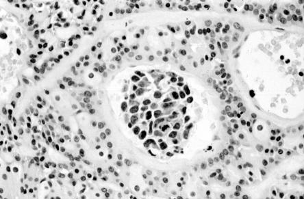
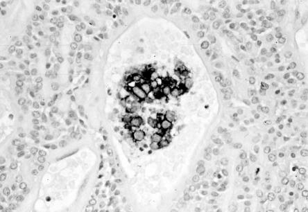
The lesion was soft, dark brown, and measured 1.5 cm maximally. Microscopic examination revealed a tumour composed of epithelial cells with abundant finely granular eosinophilic cytoplasm arranged in a predominantly microtubular architectural pattern. A minority of the tubules were cystically dilated, and many contained periodic acid Schiff positive eosinophilic secretions. Occasional goblet cells were seen. The nuclei were regular, with inconspicuous nucleoli, and the appearances were felt to be in keeping with an oncocytoma (oxyphilic adenoma). The tumour was highly vascular, and many vessels contained a distinct intravascular population of cells intermingled with a meshwork of fibrin strands, which almost occluded some vessels. These cells were large, with a moderate amount of amphophilic cytoplasm and distinctly pleomorphic nuclei (fig 1), with one to several prominent nucleoli. Immunohistochemistry showed the expression of CD45 (leucocyte common antigen), and the B cell marker CD20 (fig 2) within the atypical cells. There was no positivity for CD3, CD5, CD10, CD23, CD43, CD45RO, CD79a, or CD68 or for Epstein-Barr virus by in situ hybridisation. These findings indicated an angiotropic lymphoma of B cell type. Polymerase chain reaction studies for immunoglobulin heavy chain gene rearrangements failed to detect a clonal lymphoid population, probably because of the extremely small numbers of malignant cells present in the biopsy.
Figure 1.
The pleomorphic cytomorphology of the intravascular cells contrasts with the cytologically bland surrounding oncocytes.
Figure 2.
The B cell marker CD20 was strongly expressed within the intravascular cell groups.
DISCUSSION
Oncocytomas of the lacrimal gland are rare.1,2 It has recently been suggested that most cases classified as lacrimal oncocytoma would more accurately be designated as the lymphocyte deficient variant of Warthin’s tumour.2 A relative predominance of areas exhibiting microtubule formation or containing cystically dilated tubules has been described as characteristic of Warthin’s tumour, rather than the more solid pattern of true oncocytoma. Goblet cells are also believed to be infrequent in true oncocytomas as opposed to Warthin’s tumour. According to these criteria, the tumour in our patient could be categorised as a lymphocyte deficient Warthin’s tumour rather than a true oncocytoma.
Angiotropic lymphoma is a rare variant of non-Hodgkin’s lymphoma having a predominantly intravascular growth pattern.3 The tumour cells are characteristically present within a meshwork of fibrin strands within vessels, and although widespread dissemination may occur, cells do not circulate within the bloodstream. The unusual intravascular location may relate to a quantitative or qualitative abnormality of cell surface receptors responsible for binding to the endothelium. Expression of CD36, an antigen believed to be involved in adhesion between endothelial and other cells, but which is rarely expressed in normal lymphocytes, has been detected in one instance.4 Most cases are of B cell origin and are regarded as a subtype of diffuse large B cell lymphoma within the World Health Organisation classification.
“The tumour in our patient could be categorised as a lymphocyte deficient Warthin’s tumour rather than a true oncocytoma”
The association of lymphocytes with oncocytic epithelium in Warthin’s tumour is widely believed to result from the tumour arising from intercalary ducts within intraparotid lymph nodes. A reactive lymphocytic response to the tumour has also been postulated on the grounds that true Warthin’s tumours may also occur within other salivary glands and lacrimal glands, which have not been described as containing lymph nodes, and it has indeed been suggested that the intensity of the lymphocytic infiltrate in Warthin’s tumour is only explicable on the basis of an immune response.5 This last hypothesis might explain the relative infrequency of a Warthin’s-like lymphocytic infiltrate in lacrimal gland oncocytomas, which because of their site are usually detected at an earlier stage than their salivary gland counterparts, perhaps before the development of an immune response.5 It is also conceivable that aberrant expression of an unidentified cell surface receptor on oncocytic epithelium is responsible for “homing” of lymphoma cells to this microenvironment. The association of oncocytoma and angiotropic lymphoma in our patient is probably coincidental, although it is possible that the as yet unexplained relation between oncocytic epithelium and lymphocytes as seen in classic Warthin’s tumour may be of relevance here. This unusual juxtaposition highlights the importance of the close examination of intravascular cells in biopsy specimens, which can easily be overlooked during the histopathological reporting of surgical specimens.
Take home messages.
This is the first report of angiotropic variant of diffuse large B cell lymphoma within a benign oncocytoma of the lacrimal sac
Angiotropic lymphoma is a very rare and usually highly aggressive variant of non-Hodgkin’s lymphoma, which classically involves the central nervous system and skin, but has been described within most organs
Its occurrence within a benign neoplasm is probably coincidental, although a close association between oncocytic epithelium and normal lymphoid cells is recognised in Warthin’s tumour of salivary and lacrimal glands
REFERENCES
- 1.Biggs SL, Font RL. Oncocytic lesions of the caruncle and other ocular adnexa. Arch Ophthalmol 1977;95:474–8. [DOI] [PubMed] [Google Scholar]
- 2.Pecorella I, Garner A. Ostensible oncocytoma of the minor salivary glands. Histopathology 1997;30:264–70. [DOI] [PubMed] [Google Scholar]
- 3.Domizio P, Cotter F, Hall PA, et al. Angiotropic large cell lymphoma (ALCL): morphological, immunohistochemical and genotypic studies with analysis of previous reports. Hematol Oncol 1989;7:195–206. [DOI] [PubMed] [Google Scholar]
- 4.Muroi K, Toya K, Suzuki T, et al. Expression of CD11b, CD14 and CD36 antigens by B-cell lymphoma. Br J Haematol 1992;80:126–7. [DOI] [PubMed] [Google Scholar]
- 5.Thackray AC, Lucas RB. Tumours of the major salivary glands. In: Atlas of tumour pathology, second series, fascicle 10. Washington: Armed Forces Institute of Pathology, 40–59.


