Abstract
A 54 year old man presented with a six month history of abdominal pain. A computerised tomography scan showed a well defined intra-abdominal unilocular mass with a calcified wall just superior to the bladder. At laparotomy, pseudomyxoma peritonei was discovered, together with a midline abdominal mass adherent to the anterior abdominal wall originating from the fundus of the bladder. The specimen consisted of a cystic mass measuring 14 × 9.5 × 7 cm overall, which contained mucoid material. Histological examination revealed that the cyst was lined by mucinous epithelium, which in areas varied from having bland morphology to showing pronounced nuclear and architectural atypia. There was abundant extracellular mucin. The specimen was extensively sampled but there was no evidence of invasion. This tumour has many unusual features, namely: the absence of destructive invasion, association with pseudomyxoma peritonei, areas of dysplasia and cystadenoma, and stromal osseous metaplasia within the wall.
Keywords: adenocarcinoma, pseudomyxoma, peritonei, urachus
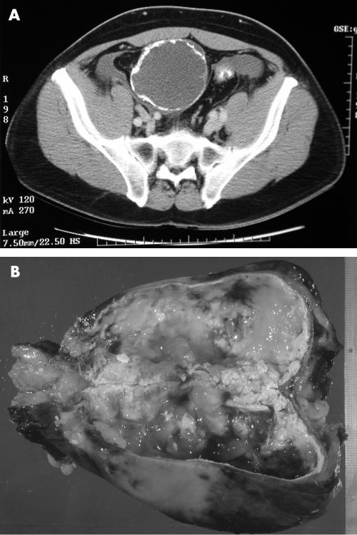
A54 year old white man presented as a referral from his general practitioner with a six month history of abdominal pain. He was suffering migratory, stabbing pains of several seconds duration and intermittent bleeding via the rectum; he had no urinary symptoms. There was no notable past medical history and no predisposing risk factors were identified. On examination, he was found to have abdominal tenderness, which was worse in the right iliac fossa, and external haemorrhoids. Biochemistry and haematology investigations were within normal limits. An ultrasound examination was undertaken, which reported a substantial cystic mass in the right iliac fossa lying below and separate from the right kidney, measuring 11 × 8 × 7 cm. Furthermore, a computerised tomography scan showed a well defined unilocular mass with a calcified wall on the anterior abdominal wall just superior to the bladder (fig 1A). Radiologically, it was thought to represent a mesenteric or enteric duplication cyst. At laparotomy, a midline abdominal mass appeared to originate from the fundus of the bladder and copious gelatinous material was present throughout the peritoneal cavity, representing a disseminated pseudomyxoma peritonei. The patient made an uneventful recovery and six months later had no recurrence.
Figure 1.
(A) Computerised tomography scan detailing a midline unilocular cystic mass just anterior to the bladder with prominent calcification within the wall. (B) Sectioning the cyst showed abundant mucoid material within the cavity.
PATHOLOGY
The specimen consisted of a cystic mass measuring 14 × 9.5 × 7 cm overall. On sectioning, it contained mucoid material (fig 1B) and calcification was noted in the wall. A small cuff of bladder wall was identified on the external surface. Occasional papillary structures projected into the cyst lumen. The cyst wall was focally ruptured.
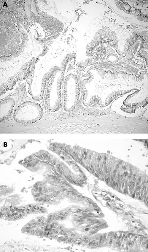
Histological examination revealed a tract within the detrusor muscle of the bladder cuff that was lined by mucinous columnar epithelium and was in continuity with the cyst. The epithelium showed changes ranging from a simple benign columnar epithelium (fig 2A) to areas of frank dysplasia, with nuclear atypia and cellular crowding and stratification (fig 2B). The architecture was also variable with flat and villous areas. The cyst wall was composed of hyalinised connective tissue with extensive foci of dystrophic calcium and an area of osseous metaplasia. Extracellular mucin was abundant and was present in the cyst wall, on the external surface, and in the detrusor wall of the bladder cuff. The specimen was extensively sampled but showed no evidence of destructive invasion.
Figure 2.
(A) Cyst wall lined by benign mucinous epithelium. (B) Epithelium showing nuclear pleomorphism, stratification, and scattered mitotic figures. Both haematoxylin and eosin stained; original magnification ×100.
DISCUSSION
Urachal cysts that arise within urachal remnants are not uncommon and, although they may be asymptomatic, they can present with urinary symptoms including mucusuria.1,2 Urachal adenocarcinoma accounts for approximately 20–30% of all bladder primary adenocarcinomas, which themselves amount to a reported 0.7–5% of all bladder tumours.3–5 In general, tumours arising in the urachus are divided into two categories—benign adenomas or malignant adenocarcinomas—resembling those of the colon and rectum. However, Paul et al have postulated that, as in colonic adenocarcinomas, there is a biological continuum.5 The case described here represents the premalignant phase and is similar to the lesion reported by Paul et al as stage 0 mucinous adenocarcinoma in situ of the urachus, although the lesion they described displayed pronounced pleomorphism, along with a high mitotic count, but lacked the abundant extracellular mucin seen in our case.5 We reported our case as “a mucinous neoplasm of uncertain malignant potential” because we felt that there were some similarities morphologically with the borderline tumour of the ovary. Urachal neoplasms can occur in urachal cysts and have been reported in association with pseudomyxoma peritonei.6,7 Another unusual feature of our present case was the presence of stromal osseous metaplasia, which has been previously described by Sasano et al.7
Take home messages.
We describe an unusual case of an in situ adenocarcinoma of the urachus arising in a giant urachal cyst, with associated pseudomyxoma peritonei
The unusual features of this tumour include: the absence of destructive invasion, association with pseudomyxoma peritonei, areas of dysplasia and cystadenoma, and stromal osseous metaplasia within the wall
The associated pseudomyxoma peritonei will probably determine the patient‘s morbidity and prognosis
“We reported our case as `a mucinous neoplasm of uncertain malignant potential‘ because we felt that there were some similarities morphologically with the borderline tumour of the ovary”
Finally, this is an unusual case of an in situ adenocarcinoma of the urachus arising in a giant urachal cyst, with associated pseudomyxoma peritonei, and it is this last feature that will probably determine the patient‘s morbidity and prognosis.
REFERENCES
- 1.Milotic F, Fuckar Z, Cicvaric T, et al. Inflamed urachal cyst containing calculi in an adult. J Clin Ultrasound 2002;30:253–5. [DOI] [PubMed] [Google Scholar]
- 2.Yagishita H, Nagayama T, Zean Z, et al. A case report of asymptomatic urachal cyst in autopsy—histopathological study of urachal cyst and review of the literature of 99 cases during a 10 year period in Japan. Hinyokika Kiyo 2001;47:849–52. [PubMed] [Google Scholar]
- 3.Cirillo RL. Urachal carcinoma. eMedicine Journal 2002;3:1. [Google Scholar]
- 4.Grignon DJ, Ro JY, Ayala AG, et al. Primary adenocarcinoma of the urinary bladder. A clinicopathologic analysis of 72 cases. Cancer 1991;67:2165–72. [DOI] [PubMed] [Google Scholar]
- 5.Paul AB, Hunt CR, Harney JM, et al. Stage 0 mucinous adenocarcinoma in situ of the urachus. J Clin Pathol 1998;51:483–4. [DOI] [PMC free article] [PubMed] [Google Scholar]
- 6.Mendeloff J, McSwain NE, Jr. Pseudomyxoma peritonei due to mucinous adenocarcinoma of the urachus. South Med J 1971;64:497–8. [DOI] [PubMed] [Google Scholar]
- 7.Sasano H, Shizawa S, Nagura H, et al. Mucinous adenocarcinoma arising in a giant urachal cyst associated with pseudomyxoma peritonei and stromal osseous metaplasia. Pathol Int 1997;47:502–5. [DOI] [PubMed] [Google Scholar]


