Abstract
A 13 year old girl presented with recurrent painful “varicosities” on her right calf. These lesions were subsequently clinically diagnosed as “cavernous haemangiomas” after normal duplex scanning and were excised. Histological examination revealed multiple glomangiomas (glomus tumours). A literature review revealed only two reported cases of nodular multiple glomangioma, so that this is the third case to be reported in the literature.
Keywords: congenital, glomangioma, glomus tumour, multiple, nodular
Glomus tumours are benign tumours of the perivasculature. They arise from modified smooth muscle cells, called glomus cells, located in the walls of the Sucquet-Hoyer canal, a specialised arteriovenous anastomosis central to thermoregulation.1 Most tumours are less than 1 cm in diameter, occurring in the dermis or subcutis in the upper and lower extremities.1 There are two forms of glomus tumour, with the more common solitary variant accounting for most of the cases (90%), and a rarer multiple variant accounting for 10% of cases2; this second form is seen most often in children and is thought to be inherited in an autosomal dominant fashion.1 The multiple variant is subdivided clinically into nodular and plaque-like lesions. Histologically, glomus tumour is composed of varying proportions of glomus cells, blood vessels, and smooth muscle.1 Depending on the predominant component, there are three variants of glomus tumour, namely: (1) angiomatoid (glomangioma) with predominant blood vessels; (2) solid (predominantly glomus cells); and (3) glomangiomyoma (predominantly smooth muscle). Multiple glomus tumours generally correspond to glomangioma.
“A literature review revealed only two reported cases of this rare variant”
In 2001, Carvalho et al reported a congenital plaque-like variant and reviewed 14 other cases. Of these 15 cases, 13 were described as the plaque-like type and two were nodular.2 In 2001, an additional case of congenital plaque-like multiple glomus tumour was reported by Lin et al.3
We report a case of congenital multiple glomangioma in a 13 year old girl, with the nodular variant. A literature review revealed only two reported cases of this rare variant.
CASE REPORT
A 13 year old girl was seen in the paediatric plastic surgery outpatient clinic in Birmingham Children’s Hospital, UK, after being referred for painful varicosities on her right calf, recurring after three previous phlebectomies (histological examination was not performed). These bluish “venous” lesions had been present since birth. The patient was experiencing pain at the site of the lesions. On standing the lesions would dilate. After each excision the lesions reappeared. The patient’s mother, grandmother, great grandmother, and great aunt each had similar lesions at different anatomical locations. Although there was no histological diagnosis for these lesions, the clinical picture supports an autosomal dominant inheritance.
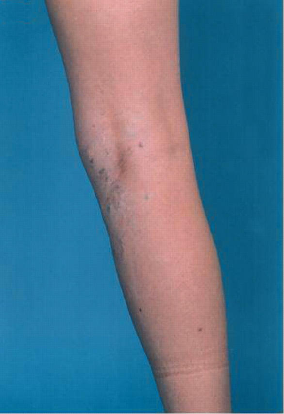
On clinical examination, there appeared to be varicosities along the long saphenous axis in her right calf (fig 1). Duplex scanning showed normal superficial and deep veins in the right leg. At this point it was assumed that the lesions were in fact scattered cavernous haemangiomata. Excision of the lesions was carried out by the vascular surgery department. At surgery, there appeared to be multiple subcutaneous haemangiomata, “like isolated knotted varicose veins”, which were excised with skin ellipses and subcutaneous tissue from the right upper part of the thigh, popliteal fossa, and medial calf. Eight lesions were excised in total.
Figure 1.

The patient’s right calf, showing varicosities along the long saphenous axis. This photograph is reproduced with the full consent of the patient.
PATHOLOGICAL FINDINGS
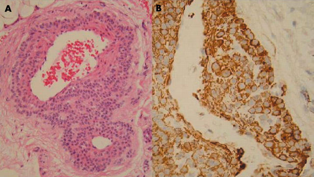
Macroscopic examination showed 10 pieces of fibro-fatty tissue, the largest of which measured 2.5 × 2.5 × 2 cm. The smallest measured 1.3 × 0.7 × 0.4 cm. Most of them showed bluish vessels. Microscopy revealed multiple diffuse lesions, present both within the dermis and deep within the subcutaneous tissue fragments, and unlikely to be excised completely. These lesions were formed of dilated vessels, with a surrounding collar of regular round cells with round nuclei and eosinophilic cytoplasm (fig 2A). There was no mitotic activity or atypia. These features were consistent with the angiomatoid variant of glomus tumour (glomangioma). The presence of the tumour within the subcutaneous tissue is in keeping with what has been described as “infiltrating glomus tumour”, which implies difficulty in complete excision and the possibility of recurrence.
Figure 2.
(A) Low power view of a lesion formed from dilated vessels, with a surrounding collar of regular round cells with round nuclei and eosinophilic cytoplasm (haematoxylin and eosin stain). (B) The tumour cells were positive for smooth muscle actin (immunohistochemical stain).
The tumour cells were immunoreactive for smooth muscle actin (fig 2B). The periodic acid Schiff stain distinctively highlighted the cytoplasmic membrane of the tumour cells. These immunohistochemical and special stains added support to the diagnosis.
DISCUSSION
Glomangioma has an early onset, with one third of cases presenting before 20 years of age.4 Familial cases have been reported with autosomal dominant transmission, incomplete penetrance, and variable expression. In 2000, Pena-Penabad et al reported two cases of familial multiple glomangiomas, but the lesions were not present from birth in the first patient and the second patient presented for consultation as an elderly adult.5 There have been several papers describing glomangiomas in the knee region,6–9 but all four papers describe a solitary tumour, which is different from the multiple type reported here. Clinically, glomangiomas appear as red to blue compressible papulonodules.2 Although solitary glomus tumours often produce pain, multiple glomangioma usually does not. In our case, the patient presented with pain. In most cases there are fewer than 10 lesions. Of the 15 cases of congenital glomangioma presenting in childhood found in the literature, 13 were described as the plaque type and two as nodular. Our patient presented in childhood with painful, bluish nodules in her right lower extremity, fitting in with the nodular multiple variant.
This is the third case in the literature of congenital multiple glomangioma, nodular type, presenting in childhood. The presence of the tumour within the subcutaneous tissue explains why it recurred.
Take home messages.
We describe a rare case of congenital multiple glomangioma, nodular type, presenting in childhood—only the third to be reported
The tumour was present within the subcutaneous tissue, causing it to recur
Supplementary Material
The patient gave full consent for the reproduction of the photograph.
REFERENCES
- 1.Fletcher C . Tumours of blood vessels and lymphatics. In: Diagnostic histopathology of tumors. Edinburgh: Churchill Livingstone 2000:75–6.
- 2.Carvalho VO, Taniguchi K, Giraldi S, et al. Congenital plaquelike glomus tumor in a child. PediatrDermatol 2001;18:223–6. [DOI] [PubMed] [Google Scholar]
- 3.Lin T-M, Tsai C-C, Tsai K-B, et al. Congenital multiple plaque-like glomangiomyoma in trunk—a case report. Kao Hsiung I Hsueh Ko Hsueh Tsa Chih 2001;17:377–80. [PubMed] [Google Scholar]
- 4.Goodman T , Abele D. Multiple glomus tumours. Arch Dermatol 1971;103:11–22. [DOI] [PubMed] [Google Scholar]
- 5.Pena-Penabad C , Garcia-Silva J, del Pozo J, et al. Two cases of segmental multiple glomangiomas in a family: type 1 or type 2 segmental manifestation? Dermatology 2000;201:65–7. [DOI] [PubMed] [Google Scholar]
- 6.Hardy P , Muller PG, Got C, et al. Glomus tumor of the fat pad. Arthroscopy 1998;14:325–8. [DOI] [PubMed] [Google Scholar]
- 7.Negri G , Schulte M, Mohr W. Glomus tumor with diffuse infiltration of the quadriceps muscle: a case report. Hum Pathol 1997;28:750–2. [DOI] [PubMed] [Google Scholar]
- 8.Mabit C , Pecout C, Arnaud JP. Glomus tumor in the patellar ligament: a case report. J Bone Joint Surg Am 1995;77:140–1. [DOI] [PubMed] [Google Scholar]
- 9.Oztekin H . Popliteal glomangioma mimicking Baker’s cyst in a 9-year-old child: an unusual location of a glomus tumor. Arthroscopy 2003;19:1–5. [DOI] [PubMed] [Google Scholar]
Associated Data
This section collects any data citations, data availability statements, or supplementary materials included in this article.


