Bronchogenic cysts are congenital anomalies evolving from the ventral foregut between the 3rd and the 7th prenatal weeks. They are lined with cuboidal or pseudostratified ciliated epithelium and may or may not be surrounded by elastic fibres, smooth muscle, and cartilage.
Bronchogenic cysts are divided into thoracic and abdominal.1 Abdominal bronchogenic cysts are rare, particularly those located exclusively within the confines of the gastric wall. Despite the fact that Gensler and colleagues1 described the first case nearly 50 years ago, only two additional cases have been reported.2,3 Recently, we identified a new case of bronchogenic cyst in the gastric mucosa. The purpose of this letter is to draw attention to an important differential diagnosis between gastric congenital intramucosal cysts and acquired intramucosal cysts also lined with ciliated cells.4
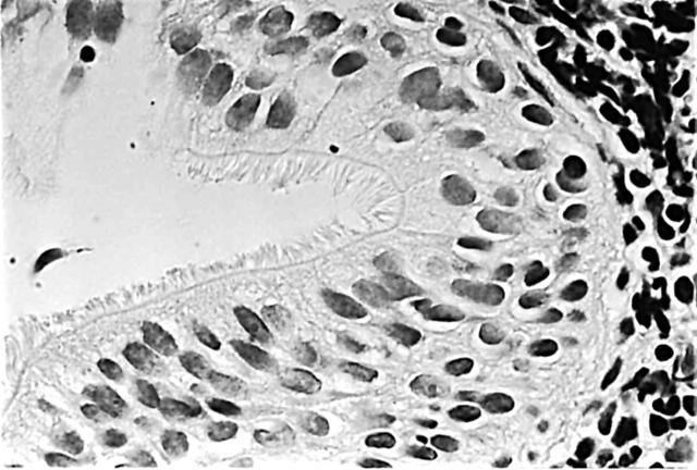
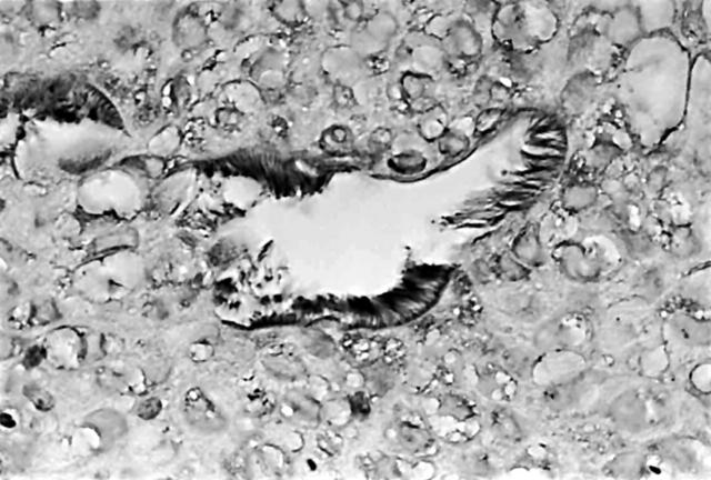
A 26 year old Swedish man presented because of periodic epigastric pain. The pain began 18 months previously and was periodically treated with proton pump inhibitors. Palpation resulted in epigastric pain. Oesophageal manometry and pH were normal. Gastroscopy showed mild oesophagitis and normal gastric mucosa. Pinch biopsies revealed grade 1 oesophagitis and normal gastric mucosa without Helicobacter pylori infection. However, an intramucosal cyst was found in one of the biopsies from the corpus. That cyst was lined with pseudostratified epithelium built from cuboidal cells (fig 1), some of them vacuolated. Cartilaginous tissue was not found but a lymphatic follicle was present in the lamina propria. The adjacent mucosa of the fundus was normal. The cells lining the luminal aspect of the cyst had densely packed cilia (fig 1). Each cilium was approximately 6 μm long and stained positively for tubulin B (fig 2). Staining with periodic acid Schiff diastase revealed only one positive clear cell. The aforementioned vacuolated cells were periodic acid Schiff negative. Staining for Ki67 (clone MIB1) showed no signs of epithelial proliferation.
Figure 1.
Gastric intramucosal bronchogenic cyst with ciliated pseudostratified epithelium (haematoxylin and eosin stain; original magnification, ×50).
Figure 2.
Gastric intramucosal bronchogenic cyst showing ciliated structures expressing tubulin B (immunohistochemical staining for tubulin B; original magnification, ×25).
The patient received omeprazol medication and the symptoms disappeared.
The histological lining of this brochogenic cyst differs from other reported intramucosal gastric cysts also lacking cartilage; namely: foveolar, fundic, pyloric, intestinal metaplastic, and ciliated metaplastic cysts.4 Cysts with ciliated metaplasia are usually located in the antrum and they are lined with a single row of gastric seromucinous cuboidal cells or with intestinal metaplastic goblet cells that have irregular cilia. Cysts with ciliated metaplasia evolve as a result of environmental factors, particularly in Asian patients harbouring a gastric carcinoma of the intestinal type.5,6
Gastric bronchogenic cysts are rare; a similar type of cyst has not been found in two large series comprising 1675 gastric biopsies7 and 3406 resected stomachs5 from patients dwelling in disparate geographical regions.
In conclusion, congenital bronchogenic cysts should be differentiated from acquired gastric ciliated cysts evolving as a consequence of environmental factors.5
References
- 1.Gensler S, Seidenberg B, Rifkin H, et al. Ciliated lined intramucosal cyst of the stomach. Ann Surg 1966;163:954–6. [DOI] [PMC free article] [PubMed] [Google Scholar]
- 2.Tanenbaum B, Levowitz B, Ponce M, et al. Respiratory choristoma of the stomach. N Y State Med J 1971;71:373–5. [PubMed] [Google Scholar]
- 3.Shireman P . Intramural cyst of the stomach. Hum Pathol 1987;18:857–8. [DOI] [PubMed] [Google Scholar]
- 4.Kato Y, Sugano H, Rubio CA. Classification of intramucosal cysts of the stomach. Histopathology 1983;7:931–8. [DOI] [PubMed] [Google Scholar]
- 5.Rubio CA, Owen D, King A, et al. Ciliated gastric metaplasia. A study of 3406 trectomy specimens from dwellers of the Atlantic and the Pacific basins. J Clin Pathol [In press.]. [DOI] [PMC free article] [PubMed]
- 6.Rubio CA, Stemmermann GN, Hayashi T. Ciliated gastric cells among Japanese living in Hawaii. Jpn J Cancer Res 1991;82:86–9. [DOI] [PMC free article] [PubMed] [Google Scholar]
- 7.Rubio CA, Jessurun J. Low frequency of intestinal metaplasia in gastric biopsies from Mexican patients: a comparison with Japanese and Swedish patients. Jpn J Cancer Res 1992;83:491–4. [DOI] [PMC free article] [PubMed] [Google Scholar]


