Abstract
Aim: To investigate the effect of myopia and myopic choroidal neovascularisation (CNV) on retrobulbar circulation in central retinal artery (CRA) and vein (CRV) and posterior ciliary artery (PCA).
Methods: 52 subjects with and without myopia were included in the study. Retrobulbar circulation was measured using colour Doppler imaging. Analysis of correlation of degree of myopia with blood flow velocity parameters was done. Circulatory differences between eyes of patients with unilateral neovascular degenerative myopia were estimated.
Results: The analysis of correlation between dioptry and blood flow velocity in the CRA, CRV, and PCA showed a significant positive correlation. Axial length was also significantly correlated with CRA and CRV blood velocity and had a tendency to be correlated with PCA blood velocity. When compared with the fellow eye, the eye with myopic CNV had significantly higher resistivity index (RI) (p=0.048) in the PCA and no significant difference in the circulatory parameters of the CRA and CRV.
Conclusion: Central retinal and posterior ciliary blood velocity decreases with the increase of the degree of myopia. PCA RI is higher in myopic CNV.
Keywords: retrobulbar circulation, myopia, choroidal neovasularisation
Degenerative myopia has been reported to be the most common cause for choroidal neovasularisation (CNV) in patients younger than 50 years of age.1 Sixty two per cent of CNV in degenerative myopia was found to be subfoveal, inflicting poor visual prognosis that poses grave social and economic consequences on patients and society.1
Choroidal and retinal circulation has been found to be decreased using colour Doppler imaging (CDI)2 and fluorescein transit times have been found to be delayed in fluorescein angiography (FAG) studies3 in degenerative myopia. However, to our knowledge, there have been no reports on the retinal and choroidal circulation in myopic patients with CNV.
In this study we aimed to investigate the correlation between the degree of myopia (dioptres and axial length), and the retinal and choroidal circulation. Furthermore, we compared the retinal and choroidal circulation in patients with unilateral myopic CNV with the intention to investigate whether circulatory disturbance may be a factor for CNV occurrence.
SUBJECTS AND METHODS
The study follows the tenets of the Declaration in Helsinki and is in accordance with the standards of the ethics committee of the University of Tokyo Hospital. All the subjects were informed about the procedure and gave their consent to participate.
Fifty two subjects were included in this study (Table 1). The subjects were randomly chosen from among patients who came for regular check ups in the general and the macular outpatient clinic at the department of ophthalmology, Tokyo University Hospital. Patients with other diseases of the eye possibly affecting the retrobulbar circulation, such as diabetic retinopathy, age related maculopathy, and glaucoma, and those with a history of laser treatment or intraocular surgery were excluded.
Table 1.
Subjects' characteristics
| Age | Sex | BPm (mm Hg) | IOP (mm Hg) | Dioptry* | AL (mm) |
| 52.2 (16.7) (range23∼78) | 27 F/25 M | 88.4 (12.9) | 14.2 (2.8) | −5.0 (6.7) (range −22∼+4.3) | 25.6 (2.7) (range 21.2∼31.6) |
Mean (SD).
BPm = mean blood pressure; IOP = intraocular pressure; *spherical equivalents; AL = axial length.
The analysis of correlation between choroidal and retinal circulation with dioptry included subjects who were recruited from among patients having cataract, and no other ophthalmic diseases and healthy volunteers. Eight patients with degenerative myopia (>−6D), who also had unilateral myopic CNV, were also included in this analysis. In the latter case, the fellow myopic eye has been used. Myopic CNV was diagnosed on the basis of fundus examination with slit lamp biomicroscopy, FAG, and indocyanine green angiography. Only one eye was included in the analysis of correlation.
Secondly, the eight patients with unilateral myopic CNV were measured in each eye and the circulatory parameters in the fellow and the eye affected with CNV were compared.
The patients underwent assessment of best corrected visual acuity after standardised refraction. Intraocular pressure (IOP) was measured by Goldmann tonometry. Brachial artery systolic (BPs) and diastolic (BPd) blood pressures were determined by an automatic blood pressure apparatus. The mean brachial artery pressure (BPm) was calculated as follows
 |
Measurements of ocular blood flow
All the measurements were performed with a CDI set (Powervision SSA-380A, Toshiba, Tokyo, Japan) using a 7 MHZ transducer. One observer (GD), masked to the subject's status, did all the measurements.
The patients were examined while in a sitting position, as described elsewhere.4 We measured the central retinal artery and vein and the posterior ciliary artery. The haemodynamic parameters measured in the aforementioned blood vessels were peak systolic blood velocity (PSV), mean velocity (MV), and end diastolic velocity (EDV). The resistivity index (RI) was calculated as follows:
 |
(1) |
The CDI method allows for accurate RI measurement because its values do not depend on the Doppler angle. We attempted to maintain the Doppler angle as parallel as possible to the measured blood vessel in order to obtain more accurate blood velocity data.
A statistical analysis for the correlation between dioptry and circulatory parameters was done by the Microsoft Excel set for data analysis. Unilateral myopic CNV patients data were analysed by the paired t test. p Values less than 0.05 were regarded as statistically significant.
RESULTS
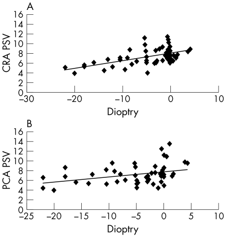
Significantly positive correlation was found between central retinal artery and vein and posterior ciliary artery blood velocity and dioptry (Table 2, Fig 1). The RI in the central retinal artery was also significantly correlated to dioptry (Table 2).
Table 2.
The correlation coefficient (r) of the circulatory parameters versus dioptry and axial length
| Dioptry | Axial length | |||
| r | p Value | r | p Value | |
| CRA | ||||
| PSV | 0.55 | <0.0001 | −0.49 | 0.0002* |
| EDV | 0.37 | 0.007 | −0.26 | 0.064 |
| MV | 0.54 | <0.0001 | −0.46 | 0.0005* |
| RI | 0.29 | 0.034 | −0.34 | 0.013* |
| CRV | ||||
| PSV | 0.34 | 0.015 | −0.23 | 0.10 |
| EDV | 0.29 | 0.040 | −0.14 | 0.34 |
| MV | 0.33 | 0.018 | −0.17 | 0.25 |
| RI | 0.12 | 0.42 | −0.12 | 0.38 |
| PCA | ||||
| PSV | 0.35 | 0.010 | −0.27 | 0.053 |
| EDV | 0.36 | 0.008 | −0.20 | 0.16 |
| MV | 0.39 | 0.003 | −0.27 | 0.054 |
| RI | 0.15 | 0.30 | −0.17 | 0.23 |
r = correlation coefficient; CRA =central retinal artery; CRV = central retinal vein; PCA = posterior ciliary artery; PSV = peak systolic velocity; EDV = end diastolic velocity; MV =mean velocity, RI = resistivity index ; *statistically significant.
Figure 1.
Correlation between dioptry and central retinal and posterior ciliary artery peak systolic velocity. CRA PSV = central retinal artery peak systolic velocity; r = 0.55, p<0.0001. PCA PSV = posterior ciliary artery peak systolic velocity; r = 0.35, p=0.01.
Axial length was significantly correlated with the PSV, MV, and RI in the central retinal artery and tended to correlate with the PSV in the central retinal vein (Table 2). In the posterior ciliary artery, the PSV and MV tended to correlate with the axial length.
There was no statistically significant difference in the dioptry and axial length between the affected and the fellow eye in unilateral myopic CNV patients (Table 3). Posterior ciliary artery RI was significantly higher in the affected eye compared to the fellow eye. None of the circulatory parameters in the central retinal artery showed any significant difference between the affected and the control eye; however, there was a tendency for a decreased PSV and EDV in the central retinal artery, and EDV and MV in the central retinal vein of the affected eye.
Table 3.
Circulatory parameters in both eyes of patients with unilateral myopic choroidal neovascularisation.
| Fellow eye | Affected eye | P value | |
| Dioptry | −12.97 (5.98) | −13.44 (3.65) | 0.76 |
| Axial length (mm) | 28.73 (2.28) | 29.11 (1.22) | 0.47 |
| CRA | |||
| PSV (cm/s) | 5.41 (0.47) | 4.96 (0.28) | 0.10 |
| EDV (cm/s) | 2.06 (0.35) | 1.88 (0.25) | 0.08 |
| MV (cm/s) | 3.31 (0.47) | 3.08 (0.12) | 0.17 |
| RI | 0.61 (0.08) | 0.61 (0.10) | 0.78 |
| CRV | |||
| PSV (cm/s) | 3.75 (0.68) | 3.44 (0.66) | 0.12 |
| EDV (cm/s) | 2.79 (0.50) | 2.48 (0.40) | 0.07 |
| MV (cm/s) | 3.18 (0.64) | 2.85 (0.48) | 0.051 |
| RI (cm/s) | 0.26 (0.06) | 0.27 (0.07) | 0.46 |
| PCA | |||
| PSV (cm/s) | 6.64 (1.87) | 7.94 (5.26) | 0.38 |
| EDV (cm/s) | 2.58 (0.61) | 2.66 (0.95) | 0.84 |
| MV (cm/s) | 4.06 (1.00) | 4.85 (2.89) | 0.41 |
| RI | 0.60 (0.05) | 0.63 (0.08) | 0.048* |
Mean (standard deviation; (n=8)
CRA- central retinal artery; CRV – central retinal vein; PCA- posterior ciliary artery; PSV- peak systolic velocity; EDV- end diastolic velocity; MV-mean velocity, RI- resistivity index; *statistically significant difference.
DISCUSSION
The present study demonstrates a significantly positive correlation between retrobulbar blood velocity parameters and the degree of myopia. These results are in agreement with a previous CDI study that analysed subjects with and without degenerative myopia in which a significantly decreased velocity in the posterior ciliary and central retinal arteries in patients with degenerative myopia has been found.2 Our results are also coherent with an FAG study that found longer transit times in patients with high and complicated myopia than in low and medium myopia, suggesting a decrease in retinal blood velocity in myopia.3
Several studies have detected a significantly negative correlation between the axial length and pulsatile ocular blood flow (POBF) and a decrease in choroidal blood flow has been suggested.5–7 Ravalico et al5 further suggested that pulse amplitude reduction might be partially related to a lower transmission of the pulsatile wave as a result of the ocular length and the degree of scleral rigidity. Our study also reveals a significantly negative correlation between the axial length and central retinal artery blood velocity and a tendency for negative correlation between posterior ciliary artery blood velocity and axial length. This may suggest that, in addition to the lower transmission of the pulsatile wave due to increased eye length, a compromised retinal and choroidal blood flow in myopia have caused a decrease of POBF in the mentioned studies. The fact that POBF studies have shown a higher correlation with axial length compared to this study could be explained not only by the pulse amplitude reduction, but also because in this study we evaluate blood velocity and not total blood flow.
Straightening of the retinal vessels, small calibre and scarce choroidal arteries, thinning and loss of choriocapillaris have been found in pathohistological studies in degenerative myopia.8 These abnormalities can be suspected to cause circulatory irregularities. The increase of ocular axial length causes stretching of scleral, choroidal, and retinal tissue. The thinning and atrophy of the choroidal and retinal tissue may decrease the need for oxygen and consequently decrease the blood circulation. One experimental study has reported that in chicks, choroidal blood flow decreased with the increase of induced ocular enlargement.9 The authors suggested that decreased choroidal blood flow is caused by either a decrease in local perfusion pressure or an increase in vascular resistivity. In our study, we did not detect an increase of the RI in uncomplicated myopia, which suggests the former possibility.
To our knowledge, there have been no reports on ocular circulation in patients having myopic CNV. As circulatory abnormalities have been found in other ophthalmic neovascular conditions, such as exudative age related maculopathy,10 we considered it important to investigate circulation in myopic CNV, as well. For that purpose we measured the retrobulbar circulation in the affected and fellow eyes of patients having unilateral myopic CNV.
The results indicating increased RI in the posterior ciliary artery of the affected eye suggests a circulatory dysfunction in the choroidal circulation. The RI is considered to represent peripheral vascular resistivity.11 Increased RI has been found in other proliferative ophthalmic diseases, such as diabetic retinopathy12 and age related maculopathy.13,14 Therefore, the fact that increased RI has been found in the eye affected with myopic CNV suggests the possibility that increased peripheral vascular resistivity could be associated with angiogenesis in degenerative myopia.
Except for the RI, other velocity parameters were not significantly different among the posterior ciliary arteries of either eyes in unilateral myopic CNV. The reason for this may be that, unlike the RI values, velocity values are dependent on the Doppler angle of measurement, which can not be always estimated with certainty in the case of the posterior ciliary artery.
In myopia, central retinal and posterior ciliary blood velocity decrease. In myopic CNV, an increase of flow resistivity in the posterior ciliary artery was found. We suggest that measures to improve choroidal circulation may be helpful in prevention of myopic CNV.
Abbreviations
CDI, colour Doppler imaging
CNV, choroidal neovascularisation
CRA, central retinal artery
CRV, central retinal vein
FAG, fluorescein angiography
PCA, posterior ciliary artery
RI, resistivity index
REFERENCES
- 1.Cohen YS, Laroche A, Leguen Y, et al. Etiology of choroidal neovascularization in young patients. Ophthalmology 1996;103:1241–44. [DOI] [PubMed] [Google Scholar]
- 2.Akyol N, Kukner S, Ozdemir T, et al Choroidal and retinal blood flow changes in degenerative myopia. Can J Ophthalmol 1996;31:113–19. [PubMed] [Google Scholar]
- 3.Avetisov ES, Savitzkaya NF. Some features in ocular microcirculation in myopia. Ann Ophthalmol 1977;9:1261–4. [PubMed] [Google Scholar]
- 4.Dimitrova G, Kato S, Tamaki Y, et al. Choroidal circulation in diabetic patients. Eye 2001;15:602–7. [DOI] [PubMed] [Google Scholar]
- 5.Ravalico G, Pastori G, Croce M, et al. Pulsatile ocular blood flow variations with axial length and refractive error. Ophthalmologica 1997;211:271–3. [DOI] [PubMed] [Google Scholar]
- 6.Mori F, Konno S, Hikichi T, et al. Factors affecting pulstile ocular blood flow in normal subjects. Br J Ophthalmol 2001;85:529–30. [DOI] [PMC free article] [PubMed] [Google Scholar]
- 7.Shih YF, Horng IH, Yang CH, et al. Ocular pulse amplitude in myopia. J Ocul Pharmacol 1991;7:83–7. [DOI] [PubMed] [Google Scholar]
- 8.Soubrane S, Coscas G, Kuhn D. Myopia. In Guyer DR, Yamuzzi LA, Chang S, et al, eds. Retina-vitreous-macula. Philadelphia: 1999:189–205.
- 9.Shih YF, Fitzgerald MEC, Reiner A. Choroidal blood flow is reduced in chicks with ocular enlargement induced by corneal incisions. Curr Eye Res 1993;12:229–37. [DOI] [PubMed] [Google Scholar]
- 10.Remulla JFC, Gaudio AR, Miller S, et al. Foveal electroretinograms and choroidal perfusion characteristics in fellow eyes of patients with unilateral neovascular age-related macular degeneration. Br J Ophthalmol 1995;79:558–61. [DOI] [PMC free article] [PubMed] [Google Scholar]
- 11.Evans W, Harris A, Danis RP, et al. Altered retrobulbar vascular reactivity in early diabetic retinopathy. Br J Ophthalmol 1997;81:279–82. [DOI] [PMC free article] [PubMed] [Google Scholar]
- 12.Guven D, Ozdemir H, Hasanreisoglu B. Hemodynamic alterations in diabetic retinopathy. Ophthalmology 1996;103:1245–9. [DOI] [PubMed] [Google Scholar]
- 13.Ciulla TA, Harris A, Chung HS, et al. Color Doppler imaging discloses reduced ocular blood flow velocities in nonexudative age-related macular degeneration. Am J Ophthalmol 1999;128:75–80. [DOI] [PubMed] [Google Scholar]
- 14.Friedman E, Krupsky S, Lane AM, et al. Ocular blood flow velocity in age-related macular degeneration. Ophthalmology 1995;102:640–6. [DOI] [PubMed] [Google Scholar]


