Measurement of the cerebrospinal fluid pressure usually requires a lumbar puncture or craniotomy to get direct access to the cerebrospinal fluid space. These techniques, however, are invasive and so carry the risk of complications such as infections and damage to the neural structures. Furthermore, owing to the leakage of cerebrospinal fluid during the puncture, the cerebrospinal fluid pressure will be altered in the moment the measurement is performed. It is, therefore, desirable to have a non-invasive method allowing the estimation of the intracerebral pressure without requiring a direct access to the brain or spinal cord. We describe a patient in whom ophthalmodynamometry strongly suggested an increased intracerebral pressure which was confirmed by eventual direct measurement.
Case report
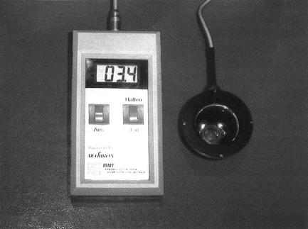
A 12 year old female patient presented with acute vomiting, massive headache, and bilateral abducens nerve palsy. Visual acuity was 20/20 in both eyes, and visual fields were unremarkable, except for an enlarged blind spot. Both optic discs showed a prominence of 0.5 mm (right eye) and 0.6 mm (left eye) as measured by confocal laser scanning tomography. Intraocular pressure measured 18 mm Hg. With topical anaesthesia, a Goldmann contact lens fitted with a pressure sensor mounted into its holding ring was put onto the cornea (Fig 1). Pressure was asserted onto the globe by slightly pressing the contact lens, and the pressure value at the time when the central retinal vein started pulsating was noted. The measurements of this new technique of ophthalmodynamometry were repeated nine times in both eyes.
Figure 1.
Photograph showing the Goldmann contact lens with a pressure sensor mounted into the holding ring of the contact lens and connected to a display.
The central retinal vein collapse pressure as the sum of the ophthalmodynamometric value plus the intraocular pressure, measured 103 relative units right eye and 98 relative units left eye. These values were significantly higher than normal values (6.1 (SD 8.4) relative units) determined previously in normal subjects (own data). Direct measurement of cerebrospinal fluid pressure by lumbar puncture performed about 5 hours later revealed a value of 107 cm water column (equivalent to 82.3 mm Hg). In combination with other clinical findings, the diagnosis of pseudotumour cerebri was made.
Comment
The central retinal vein is the only structure whose appearance depends on its inner pressure, and which runs through the cerebrospinal fluid space and which is accessible from outside the body without any invasive procedure being performed. After exiting the eye through the optic disc, the central retinal vein goes through the retrobulbar part of the optic nerve before it traverses the subarachnoidal and subdural spaces of the optic nerve and pierces the optic nerve meninges. The pressure in the central retinal vein is thus at least as high as the cerebrospinal fluid pressure. The central retinal vein collapse pressure may be measurable by ophthalmodynamometry since the vein will start to pulsate, if the sum of intraocular pressure plus an external pressure exerted onto the eye equals the diastolic pressure of the central retinal vein.1–4 The intraocular pressure can be determined by applanation tonometry, and the additional pressure exerted onto the globe can be measured by the ophthalmodynamometer. In the ophthalmodynamometers used in the 1960s and 1970s, determinations of the central retinal vein pressure were often difficult or almost impossible so that the central retinal vein pressure has usually not been measured.5 The new ophthalmodynamometer used in the present study (Fig 1) may overcome some of the problems associated with the old ophthalmodynamometers. In a previous study on the reproducibility of the new technique, the variation of the central retinal vein collapse pressure was 15.9% (SD 11.9%). The present study suggests that, in patients with markedly increased intracerebral pressure, the new, Goldmann lens associated, ophthalmodynamometer may provide information about the intracerebral pressure by estimating the central retinal vein collapse pressure. It may be helpful for the neuro-ophthalmological diagnosis of diseases associated with increased intracerebral pressure.
Proprietary interest: none.
References
- 1.Meyer-Schwickerath R, Kleinwachter T, Firsching R, et al. Central retinal venous outflow pressure. Graefes Arch Clin Exp Ophthalmol 1995;233:783–8. [DOI] [PubMed] [Google Scholar]
- 2.Morgan WH, Yu DY, Cooper RL, et al. Retinal artery and vein pressures in the dog and their relationship to aortic, intraocular and cerebrospinal fluid pressures. Microvasc Res 1997;53:211–21. [DOI] [PubMed] [Google Scholar]
- 3.Firsching R, Schutze M, Motschmann M, et al. Venous opthalmodynamometry: a noninvasive method for assessment of intracranial pressure. J Neurosurg 2000;93:33–6. [DOI] [PubMed] [Google Scholar]
- 4.Draeger J, Rumberger E, Hechler B. Intracranial pressure in microgravity conditions: non-invasive assessment by ophthalmodynamometry. Aviat Space Environ Med 1999;70:1227–9. [PubMed] [Google Scholar]
- 5.Wunsh SE. Ophthalmodynamometry. N Engl J Med 1969;281:446. [PubMed] [Google Scholar]
- 6.Jonas JB. Reproducibility of ophthalmodynamometric measurements of the central retinal artery and vein collapse pressure. Br J Ophthalmol 2003 (in press). [DOI] [PMC free article] [PubMed]


