Keratoconus is an idiopathic, progressive, non-inflammatory ectasia of the axial cornea. Its association of other systemic disorders or ocular disease have been reported, but its specific origin remains unknown. Recently, Munier and associates detected that four types of autosomal dominant corneal dystrophy result from mutation in the human transforming growth factor β induced gene (βigH3), the product of which has shown to be the protein keratoepithelin (R555W for granular corneal dystrophy, R555Q mutation for Reis-Bückler’s corneal dystrophy, R124C mutation for lattice corneal dystrophy type I, and R124H mutation for Avellino corneal dystrophy).1 Molecular genetic analysis of various corneal dystrophies, which had previously presented an insuperable challenge to clinical diagnosis, now clearly demonstrates the distinct phenotypes.2 We report a rare case of bilateral keratoconus in association with Avellino corneal dystrophy diagnosed by molecular genetic analysis.
Case report
A 35 year old man had complained blurred vision in both eyes for several years. His general health was good and there was no history of atopic disease, connective tissue disease, or ocular trauma. His familial history was unknown.
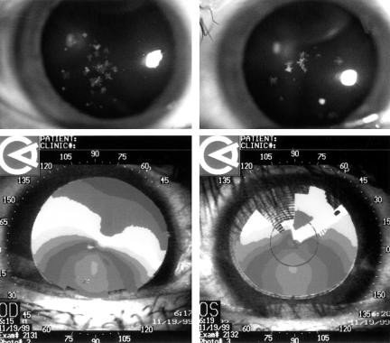
His best corrected visual acuity was RE 20/50 and LE 20/100. Slit lamp examination revealed bilateral non-inflammatory corneal thinning with protrusion of the central thinning areas. Fleischer ring was found in both corneas. Central corneal thickness was 428 μm on the right and 421 μm on the left measured by ultrasonic pachymetry. There was also clinical evidence of granular corneal dystrophy in both eyes. Discrete grey-white opacities and star-shaped spicular opacities were seen in anterior stroma (Fig 1, top). Computed corneal topography showed inferior steeping consistent with the diagnosis of keratoconus (Fig 1, bottom). With rigid gas permeable contact lenses his visual acuity corrected to 20/20 right and 20/25 left. The remainder of the ocular examination was unremarkable.
Figure 1.
Slit lamp photographs RE (top left) and LE (top right) show discrete grey-white opacities and star-shaped spicular opacities in anterior stroma. (Bottom left and right) Computed corneal topography shows inferior steepening resulting in the diagnosis of keratoconus.
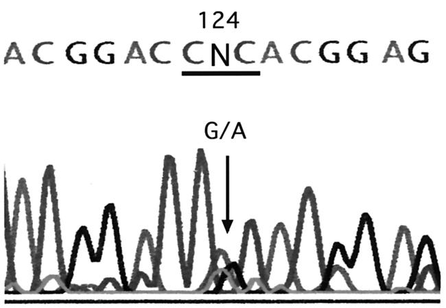
After obtaining informed consent, we collected venous blood from the patient and extracted genomic DNA. Using appropriate primers,1 we amplified exons 4 and 12 of the βigH3 gene by polymerase chain reaction (PCR) and directly sequenced the products. We detected a heterozygous G→A transition in codon 124 that results in a substitution from arginine to histidine in this patient (Fig 2). These genetic findings were consistent with Avellino corneal dystrophy.
Figure 2.

Results of direct sequencing analysis of the exon 4 of βigH3 gene. Heterozygous G→A transition is seen at the second position of codon 124 (arrow).
Comment
To our knowledge, this is the first molecular genetic report of a bilateral association of keratoconus with Avellino corneal dystrophy. There is only one case report in the literature of a patient with keratoconus associated with Avellino corneal dystrophy. Sassani and associates reported the bilateral association of keratoconus and Avellino corneal dystrophy, which was diagnosed histopathologically.3 On the other hand, there are five reports with keratoconus associated with granular corneal dystrophy.4–8 However, those cases were diagnosed clinically, not histopathologically or genetically.4–7 A clinical diagnosis of the different types of corneal stromal dystrophy is difficult, especially for granular corneal dystrophy and Avellino corneal dystrophy.9 Some cases previously reported as granular corneal dystrophy might be actually cases of Avellino corneal dystrophy.
The involvement of genetic factors has been reported in keratoconus, but its hereditary pattern was not identified. A gene for at least one form of hereditary keratoconus has been mapped to human chromosome 21.10 In our case, it is unclear whether a genetic factor had a role in the simultaneous development of keratoconus and Avellino dystrophy. There may be some linkage between the genes responsible for these two abnormalities. In our case, molecular genetic analysis clearly demonstrated the presence of distinct phenotype, which had not previously presented clinically.
The authors have no proprietary interest in any aspects of this work.
References
- 1.Munier FL, Korvastska E, Djemai A, et al. Kerato-epithelin mutations in four 5q31-linked corneal dystrophies. Nat Genet 1997;15:247–51. [DOI] [PubMed] [Google Scholar]
- 2.Klintworth GK. Advances in the molecular genetics of corneal dystrophies. Am J Ophthalmol 1999;128:747–54. [DOI] [PubMed] [Google Scholar]
- 3.Sassani JW, Smith SG, Rabinowitz YS. Keratoconus and bilateral lattice-granular corneal dystrophies. Cornea 1992;11:343–50. [DOI] [PubMed] [Google Scholar]
- 4.Yoshida H, Funabashi M, Kanai A. Histological study of the corneal granular dystrophy complicated by keratoconus. Folia Ophthalmol Jpn 1980;31:218–23. [Google Scholar]
- 5.Koomoto R, Koomoto M, Moriyama H. One case of granular corneal dystrophy associated keratoconus and esotropia. Folia Ophthalmol Jpn 1984;35:2002–7. [Google Scholar]
- 6.Vajpayee RB, Snibson GR, Taylor HR. Association of keratoconus with granular corneal dystrophy. Aust NZ J Ophthalmol 1996;24:369–71. [DOI] [PubMed] [Google Scholar]
- 7.Mitsui M, Sakimoto T, Sawa M, et al. Familial case of keratoconus with corneal granular dystrophy. Jpn J Ophthalmol 1998;42:385–8. [DOI] [PubMed] [Google Scholar]
- 8.Wollensak G, Green WR, Temprano J. Keratoconus associated with corneal granular dytrophy in a patient of Italian origin. Cornea 2002;21:121–2. [DOI] [PubMed] [Google Scholar]
- 9.Kocak-Altintas AG, Kocak-Midillioglu I, Akarsu AN, et al. BIGH3 gene analysis in the differential diagnosis of corneal dystrophies. Cornea 2001;20:64–8. [DOI] [PubMed] [Google Scholar]
- 10.Rabinowitz YS, Zu L, Yang H, et al. Keratoconus: non-parametric linkage analysis suggests a gene locus near the centromere of chromosome 21. Invest Ophthalmol Vis Sci 1999;40:S564. [Google Scholar]


