Birdshot chorioretinopathy (BSCR) is a rare inflammatory disease, which generally follows a chronic course of progressive loss of vision to 6/60 or worse.1 However, the vision may stabilise1 or, rarely, improve slightly.2 Commonly prescribed treatment regimens, including oral steroids, and one or two immunosuppressive agents, may stabilise but generally do not cure the condition.3
We describe a case of BSCR in which there was a marked improvement in Snellen acuity and retinal function, measured with wide field multifocal electroretinography (WF-mfERG),4,5 in a patient intolerant of conventional treatment, who self medicated with an antioxidant preparation.
CASE REPORT
A 53 year old white woman was referred with a 2 year history of decreased vision and floaters affecting both eyes. Snellen acuity was 6/24, N8 in the right eye and 6/12, N8 in the left eye. Moderate vitritis, cystoid macular oedema, swelling of the optic discs, and the presence of scattered, deep, pale creamy white lesions, were compatible with a diagnosis of BSCR, supported by positive HLA-A29 serology.
A reducing dose of oral prednisolone in conjunction with cyclosporin was commenced. There was no improvement in visual symptoms and, owing to the development of cellulitis around the umbilicus, cyclosporin was stopped.
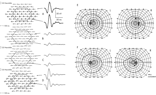
She was referred to the regional uveitis service. WF-mfERG was performed at presentation, using a custom built system comprising a 61 hexagonal array.4 This test facilitates localised assessment of retinal function, within a 90° retinal field. WF-mfERG showed a global reduction of P1 amplitude responses in both eyes (Fig 1B). A lower dose of cyclosporin was tried but subsequently discontinued because of significant gastrointestinal symptoms.
Figure 1.
Multifocal electroretinogram (mfERG) of the patient’s right eye compared to a normal response. (A) Shows a normal mfERG; the central and peripheral responses are grouped and averaged. The normal range of the central response is 74–122 nV and the normal range of the peripheral response is 61–108 nV. The normal range for mfERG implicit times is 32–42 ms. The ranges are the 5% and 95% confidence limits derived from 50 age-matched controls. (B) Shows the mfERG of the patient’s right eye performed in 1999 at presentation to the regional uveitis clinic. The average central response is 30 nV and the average peripheral response is 31 nV. (C) Shows the mfERG of the right eye performed 1 year later. The average central response is reduced to 21 nV and the average peripheral response is 30 nV. The implicit time is delayed by 12–54 ms. (D) Shows the mfERG of the patient’s right eye performed 1 year later, 6 months after starting the antioxidant preparation. The average central response shows a marked improvement to 95 nV and the average peripheral has improved to 83 nV. The implicit time delay has also improved by 6 ms to 48 ms. (E) Goldmann perimetry findings before commencing antioxidant treatment. (F) Goldmann perimetry findings 5 months after commencing antioxidant treatment.
Over the next 18 months, tacrolimus, azathioprine, and mycophenolate were all prescribed separately in conjunction with a maintenance dose of 5 mg of prednisolone. Tacrolimus 1 mg twice daily was discontinued after 2 weeks because of intolerable side effects. Treatment with azathioprine 50 mg three times daily continued for 7 months with no improvement in vision. Mycophenolate 1 mg twice daily was stopped at the patient’s request after a year, owing to progressive deterioration of vision and a general feeling of malaise. In addition, prednisolone was tailed off and stopped after 20 months of steroid treatment. The visual acuity at this stage was 6/36, N14 and 6/12, N14, and the patient was taking no conventional medication. The amplitude of the central and peripheral WF-mfERG responses recorded a year after presentation had deteriorated further (Fig 1C).
Three months after discontinuing all conventional treatment, the patient began to self medicate with an antioxidant preparation, the contents of which included white and maritime pine bark, grape seed extract, β carotene, and vitamins C and E (Table 1). After 6 months, Snellen acuity had improved to 6/15, N10 in the right eye and 6/9, N8 in the left. In addition, the amplitude of the central WF-mfERG responses had improved from 21 nV to 95 nV in the right eye and from 35 nV to 167 nV in the left eye (normal range 74–122 nV). The amplitude and implicit time of the average peripheral WF-mfERG responses had also improved in both eyes (Fig 1D).
Table 1.
List of ingredients in antioxidant preparation. The amount per serving was specified only for those three ingredients listed
| Ingredient | Amount per serving | Percentage of recommended daily intake |
| Astaxanthin | ||
| Grape seed extract | ||
| Decaffeinated green tea extract | ||
| Tumeric extract | ||
| Rosemary officinalis extract | ||
| Alpha lipoic acid | ||
| Ellagic acid | ||
| Resveratrol | ||
| Bioflavonoid complex | ||
| Carotenoid complex | 9500 IU | 190% |
| Co-enzyme Q-10 | ||
| N-acetyl cysteine | ||
| Esterified vitamin C | 82 mg | 137% |
| Vitamin E | 14 IU | 47% |
| Glutathione | ||
| Quercitin | ||
| White pine bark extract | ||
| Taurine | ||
| Inositol | ||
| Potassium sulphate | ||
| Selenium | ||
| Selenomethione | ||
| Copper | ||
| Zinc monomethionine |
She continued on the antioxidant regimen and 6 months later, the visual acuity and retinal function in both eyes, recorded with the WF-mfERG, had remained stable.
COMMENT
WF-mfERG investigations showed a marked improvement in visual function of both eyes after commencing the antioxidant preparation. This observation mirrored the recovery of Snellen acuity but did not correlate with Goldmann perimetry findings (Fig 1E and F).
Antioxidants are thought to scavenge free radicals produced in the retina following light absorption, and thus prevent cellular damage that they may produce.6,7 The pathophysiology of BSCR is unclear; therefore the role of antioxidants in reversing the damage caused by BSCR is purely speculative.
In this case it is unclear whether the improvement in visual function, recorded with WF-mfERG was spontaneous, whether it was as a late result of previous conventional treatment, or whether it was secondary to the commencement of a complex non-prescribed antioxidant preparation. The role of antioxidant treatment in BSCR needs further appraisal, perhaps using the HLA-A29 transgenic mouse, which has been shown to spontaneously develop a retinopathy that is histologically very similar to that found in BSCR.8
References
- 1.Ryan SJ, Maumenee AE. Birdshot retinochoroidopathy. Am J Ophthalmol 1980;89:31–45. [DOI] [PubMed] [Google Scholar]
- 2.Priem HA, Oosterhuis JA. Birdshot chorioretinopathy: clinical characteristics and evolution. Br J Ophthalmol 1988;72:646–59. [DOI] [PMC free article] [PubMed] [Google Scholar]
- 3.Bloch-Michel E, Frau E. Birdshot retinochorioretinopathy and HLA-A29+ and HLA-A29 idiopathic retinal vasculitis: comparative study of 56 cases. Can J Ophthalmol 1991;26:361–6. [PubMed] [Google Scholar]
- 4.Parks S, Keating D, Evans AL. Wide field functional imaging of the retina. IEEE Medical Applications of Signal Processing 1999;99/107:9/1–9/6. [Google Scholar]
- 5.Sutter EE, Tran D. The field topography of ERG component in man—1. The photopic luminance response. Vis Res 1992;32:433–46. [DOI] [PubMed] [Google Scholar]
- 6.AREDS. A randomised, placebo-controlled, clinical trial of high-dose supplementation with Vitamins C and E, beta-carotene, and zinc for age-related macular degeneration and vision loss. AREDS Report No 8. Arch Ophthalmol 2001;119:1417–36. [DOI] [PMC free article] [PubMed] [Google Scholar]
- 7.Jacques PF, Chylack LT Jr, Hankinson SE, et al. Long-term nutrient intake and early age-related nuclear lens opacities. Arch Ophthalmol 2001;119:1009–19. [DOI] [PubMed] [Google Scholar]
- 8.Szpak Y, Vieville J-C, Tabary T, et al. Spontaneous retinopathy in HLA-A29 transgenic mice. Prot Natl Acad Sci 2001;98:2572–6. [DOI] [PMC free article] [PubMed] [Google Scholar]


