The chromosome 22q11.2 deletion syndrome (22q11DS) encompasses velocardiofacial syndrome (VCFS), DiGeorge syndrome (DGS), and conotruncal anomaly face syndrome (CTFS) and is the result of a microdeletion of chromosome band 22q11.2. It is a relatively common genetic anomaly estimated to occur in approximately one in 4000 live births. The 22q11.2 deletion can arise de novo or can have an autosomal dominant inheritance. The condition is thought to be due in part to abnormal development of the pharyngeal arch structures. Clinical findings are extensive and highly variable between patients. Prominent features include cardiac defects, cleft palate, dysmorphic facies, maldescent of the thymus, hypoparathyroidism, immune deficiency and developmental delay. Ocular findings include hypertelorism,1 retinal vascular tortuousity, narrow palpebral fissures, small optic nerves, iris nodules, cataracts,2 and iris coloboma.3 We present a case of a boy who was found to have bilateral disc swelling that led to a diagnosis of 22q11DS.
Case report
A 14 year old boy presented to the accident and emergency department after having a generalised seizure. He had been admitted to another hospital, 2 days before this, with a sudden onset of collapse and subsequent respiratory arrest. At that time he was noted to have swollen optic discs and a head computed tomography scan done there was reported as normal. He had further seizures after admission to our hospital. Blood testing revealed low plasma calcium and high plasma phosphate levels. The patient had been complaining of back pain in recent months and his mother said that he had shrunk by a couple of centimetres over the past year. She also said that he had always been clumsy and he had been diagnosed as dyslexic at the age of 7. He had a history of behavioural problems which his family said often settled during holidays in the sun. A thoracic spine x ray revealed wedge-shaped fractures of three vertebral bodies. Further blood testing showed a relatively low parathormone level confirming the diagnosis of hypoparathyroidism. He was then referred to the ophthalmology department for assessment of his disc swelling.
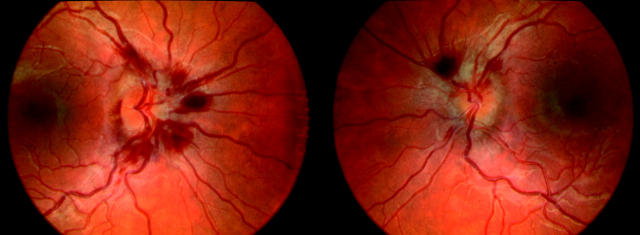
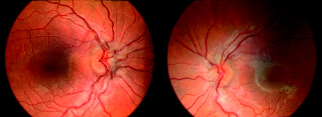
He had been seen in the eye clinic a year before this presentation complaining of coloured lines in his field of vision. No abnormality was found and his discs on that occasion were noted to be normal. On examination this time he was noticed to have dysmorphic features, notably palpebral fissures slanting medially upwards, and abnormally formed low set ears. Visual acuities were 6/5 in both eyes, colour vision testing was unremarkable, and there was no relative afferent pupillary defect. There was no sign of cataract formation or any other abnormality of the anterior segments. Funduscopy revealed bilateral disc swelling with extensive peripapillary haemorrhages in both eyes (fig 1). The diagnosis of hypoparathyroidism combined with the patient’s abnormal facies raised the suspicion of a genetic disorder and blood was then sent for chromosomal analysis. His hypoparathroidism was treated with vitamin D and calcium supplements and he responded well with his calcium reaching normal levels within a few days. Examination 6 weeks after his calcium had normalised showed most of the haemorrhages and disc swelling had cleared (fig 2). Results of chromosomal testing revealed a region 11.2 microdeletion of the long arm (q) of chromosome 22. Other members of his family are now also being tested.
Figure 1.
Shows bilateral disc swelling with extensive peripapillary haemorrhages in both eyes.
Figure 2.
Examination 6 weeks later showed most of the haemorrhages and disc swelling had cleared.
Comment
Chromosome 22q11.2 deletion syndrome is one of the more common causes of congenital and childhood hypoparathyroidism which can lead to hypocalcaemia. Hypocalcaemia is a known cause of disc swelling,4 the mechanism of which is not known. Some patients with hypocalcaemic disc swelling have a loss of acuity and field typical of an optic neuropathy, while in others the features resemble papilloedema, with no significant visual loss.5 The swelling usually resolves after the calcium level returns to normal.6 22q11DS is probably underdiagnosed. This case illustrates the importance of a correct and early diagnosis of this relatively common genetic disorder so that treatment can begin in an effort to prevent further medical and developmental complications. The highly variable clinical features require a high level of awareness of the condition across several different disciplines. Patients, especially children, presenting with swollen optic discs and who have normal imaging studies of the brain should have a calcium level checked. If abnormal and it is found to be due to hypoparathryoidism then chromosomal analysis should be considered, especially if other parts of the history or examination raise the suspicion of a genetic disorder.
References
- 1.Kitano I, Park S, Kato K, et al. Craniofacial morphology of conotruncal anomaly face syndrome. Cleft Palate Craniofac J 1997;34:425–9. [DOI] [PubMed] [Google Scholar]
- 2.Mansour AM, Goldberg RB, Wang FM, et al. Ocular findings in the velo-cardio-facial syndrome. J Pediatr Ophthalmol Strabismus 1987;24:263–6. [DOI] [PubMed] [Google Scholar]
- 3.Morrison DA, Fitzpatrick DR, Fleck BW. Iris coloboma and a microdeletion of chromosome 22: del(22)(q11.2). Br J Ophthalmol 2002;86:1316. [DOI] [PMC free article] [PubMed] [Google Scholar]
- 4.Walsh FB, Murray R. Ocular manifestations of disturbances in calcium metabolism. Am J Ophthalmol 1953;36:1657–76. [DOI] [PubMed] [Google Scholar]
- 5.McLean C, Lobo R, Brazier JD. Optic disc involvement in hypocalcaemia with hypoparathyroidism: papilloedema or optic neuropathy? Neuro-Ophthalmology 1998;20:117–24. [Google Scholar]
- 6.Kent CJ, Jakobiec FA. Ophthalmic manifestations of some metabolic and endocrine disorders. In: Albert DM, Jakobiec FA, eds. Principles and practice of ophthalmology, 2nd ed. Philadelphia: Saunders, 2000:4730–42.


