Abstract
Background/aims: To study the optic nerve head (ONH) characteristics in a cross sectional study with confocal laser scanning tomography using the Heidelberg retina tomograph (HRT I) and thereby to obtain a new HRT database for comparison of healthy and glaucomatous eyes.
Methods: White adults with no history of ocular pathology were eligible for the study. The examination comprised: assessment of visual acuity; slit lamp examination of the anterior and posterior segment; Goldmann applanation tonometry; computerised perimetry, and optic nerve head tomography with HRT. Eyes with ocular pathology were excluded. Mean (standard deviation, SD) and difference between right and left eye (RE–LE) were calculated for HRT I measurements. Differences in mean topographic parameters between male and female participants and between the age quartiles were analysed. The study included 1764 eyes of 882 healthy adults (154 females and 728 males, mean age of 46.8 (SD 8.6) years). The population investigated was larger and older in comparison with similar studies using confocal laser scanning tomography.
Results: With HRT I, a mean disc area of 1.82 (SD 0.39) mm2, a mean cup area of 0.44 (SD 0.32) mm2 and a mean cup:disc area ratio of 0.22 (SD 0.13) was observed. Right eyes showed a larger mean retinal nerve fibre layer thickness (RNFLT) (0.263 (SD 0.066) mm) compared with left eyes (0.252 (SD 0.065) mm, p<0.001). Higher values in younger volunteers (mean age 35.7 years) in comparison with elderly participants (mean age 59.1 years) were noted for disc area (1.84 mm2 v 1.78 mm2) and mean RNFLT (0.263 (SD 0.06) mm v 0.249 (SD 0.07) mm) but were not significant (p>0.01). The presented results differ from published data on ONH measurements of healthy volunteers with different techniques.
Conclusion: The observed differences in ONH measurements between left and right eyes seem not to be of clinical importance. This is also true for age or sex dependent changes in ONH topographies. The presented data provide a new basis for comparison of optic disc characteristics between healthy eyes and glaucomatous eyes.
Morphology of the optic nerve head (ONH) is of importance in the diagnosis and follow up of glaucoma. 1, 2 The laser scanning ophthalmoscope permits to analyse the optic disc topography of the ONH, thereby detecting glaucomatous as well as other changes of the optic disc. 3– 5 To evaluate glaucomatous eyes it is necessary to obtain reliable comparative data and study the topographic morphology of the ONH in normal eyes. The normalised rim:disc area ratio may be useful for glaucoma screening, diagnosis, and follow up. The calculation of this parameter relies on a comparison database with measurements obtained from 100 healthy individuals with a mean age of 36 years. 4, 6 The mean age of patients with primary open angle glaucoma is higher and ophthalmoscopy shows a broad variability of healthy ONHs. A larger comparison database with older participants would result in a better interpretation of topographic ONH measurements regarding glaucoma.
Previous studies investigated the influence of age, refractive error, optic disc size, intraocular pressure, and other optic disc parameters in normal eyes. 7– 10 As shown in table 4 , the results of these studies differ. To further elucidate the optic nerve head characteristics among elderly individuals with normal eyes, ONHs of a larger healthy population were analysed with confocal laser scanning tomography.
Table 4.
Comparison between the published literature and our findings for optic nerve head measurements by HRT I in normal eyes. Results are shown as mean (SD)
| Optic nerve head measurements by HRT I in normal eyes | |||||
| Ghergel et al 7 | Bartz-Schmidt et al 4 | Iester et al 9 | Nakamura et al 18 | Presented data | |
| Number of participants | 157 | 100 | 62 | 77 | 882 |
| Mean age, years (range) | 47.8 (14–77) | 36 (9–67) | 48.9 (NA) | 56 (21–84) | 46.8 (35–70) |
| HRT I software version | 1.12 | 1.11 | 1.11 | 1.11 | 2.01 |
| Disc area (mm2) | |||||
| RE | 1.92 (0.38) | 2.14 (0.48)† | 2.47 (0.67)† | 2.15 (0.50)‡ | 1.83 (0.39) |
| LE | 1.90 (0.37) | 1.81 (0.40) | |||
| Cup area (mm2) | |||||
| RE | 0.49 (0.26) | 0.63 (0.42)† | 0.73 (0.46)† | 0.55 (0.42)‡ | 0.44 (0.32) |
| LE | 0.47 (0.28) | 0.44 (0.32) | |||
| Cup:disc area ratio | |||||
| RE | 0.24 (0.10) | NA | 0.28 (0.14)† | 0.24 (0.14)‡ | 0.22 (0.13) |
| LE | 0.23 (0.11) | 0.22 (0.13) | |||
| Rim area (mm2) | |||||
| RE | 1.43 (0.28) | 1.51 (0.37)† | 1.76 (0.53)† | 1.59 (0.34)‡ | 1.39 (0.27) |
| LE | 1.42 (0.25) | 1.37 (0.27) | |||
| Mean RNFLT (mm)* | |||||
| RE | 0.245 (0.06) | NA | 0.26 (0.10)† | 0.250 (0.080)‡ | 0.263 (0.066) |
| LE | 0.270 (0.06) | 0.252 (0.065) | |||
| Rim volume (mm3) | |||||
| RE | 0.37 (0.12) | NA | 0.49 (0.29)† | 0.44 (0.15)‡ | 0.38 (0.13) |
| LE | 0.40 (0.13) | 0.36 (0.12) | |||
*Mean retinal nerve fibre layer thickness (HRT).
†Randomised selection of measured eyes.
‡Means for both eyes not available.
NA, not available.
METHODS
A total of 882 healthy white adults were examined between July 1998 and October 2000. Inclusion criteria for participants were: white adult, aged 35 to 70 years; no ocular pathology; no optic disc abnormalities; no ocular surgery, ocular trauma; no neurological disease; no intraocular pressure >21 mm Hg, and no visual field abnormalities.
The standardised examination of both eyes included: assessment of best corrected visual acuity; slit lamp examination of the anterior and posterior segment; keratometry with a Schwind 90 Ophthalmometer (Herbert Schwind GmbH, Kleinostheim, Germany); confocal laser scanning tomography with the Heidelberg retina tomograph (HRT) I (Heidelberg Engineering GmbH, Heidelberg, Germany); computerised 30° field perimetry (OCTOPUS 500 EZ; Interzeag AG, Schlieren, Switzerland), and Goldmann applanation tonometry.
ONH imaging with the HRT I was performed using mean topographies based on three series of HRT images (256×256 pixels), scan angle of 10°. For calculations of optic disc parameters with HRT software version 2.01 the standard reference plane was placed 50 µm posterior to the mean height of the contour line defining the disc margin in a temporal segment between 350° and 356°, as described in the literature. 11 On the topographic images, the optic disc margin was outlined along the inner margin of the scleral ring of Elschnig by one investigator and then independently reviewed for accuracy by two other investigators.
Glaucoma was excluded in all individuals in accordance with the guidelines of the European Glaucoma Society. 12 Optic discs with oblique insertion, as well as small and large papillae were included.
HRT data were transferred to SPSS statistical software version 10.0 (SPSS Inc, Chicago, IL, USA) for further analysis. 13 Measurements of disc area, rim area mean retinal nerve fibre layer thickness (RNFLT), and rim volume were tested for normal distribution. Right and left eyes were analysed separately. The data analysis focused disc area, cup area, cup to disc area ratio, rim area, rim volum, and mean RNFLT. Differences between right and left eyes, men and women, and age groups (age quartiles) were calculated and tested for significance. Differences in means of topographic parameters were evaluated with non-parametric tests (Wilcoxon test). The statistical analysis was performed at the Department for Medical Statistics, Informatics and Epidemiology, University of Cologne, Germany.
The statistical analysis included 1764 eyes of 882 healthy adults; 154 female and 728 male; mean age 46.8 (SD 8.6) years, range 35–70 years. The mean (SD) refractive error was +0.04 (1.91) D, range: −10.75 D to +10 D and a mean astigmatism of −0.47 (SD 0.77) D, range: 0 D to −5.75 D was observed.
RESULTS
Table 1 shows the mean values of the right (RE) and left eyes (LE) for optic disc topographic parameters as measured by HRT I with standard deviation (SD), median, and range values. Additionally, this table shows the mean for the difference between right and left eyes of the topographic parameters and the statistical significance (Wilcoxon test). Statistically significant differences (RE–LE) of 1764 healthy eyes of adults were found for rim volume and mean RNFLT (p<0.001). Pearson’s correlation coefficients of disc area with cup area were 0.72 (RE) and 0.75 (LE) (p<0.01) and of disc area with rim area were 0.60 (RE) and 0.59 (LE) (p<0.01) respectively.
Table 1.
Mean (SD) optic disc measurements by HRT I in normal eyes, median, range, and mean (SD) individual difference between right and left eye (RE–LE) and significance (Wilcoxon test)
| 882 participants, 1764 normal eyes | ||||||||
| Right eyes | Left eyes | Difference RE–LE | ||||||
| Mean (SD) | Median | Range | Mean (SD) | Median | Range | Mean (SD) | p Value | |
| Disc area (mm2) | 1.83 (0.39) | 1.80 | 0.64–3.25 | 1.81 (0.39) | 1.77 | 0.88–3.77 | +0.017 (0.295) | 0.04 |
| Cup area (mm2) | 0.44 (0.32) | 0.39 | 0–1.80 | 0.44 (0.32) | 0.37 | 0–1.84 | +0.004 (0.204) | 0.64 |
| Cup:disc area ratio | 0.22 (0.13) | 0.23 | 0–0.69 | 0.22 (0.13) | 0.22 | 0–0.69 | +0.0001 (0.094) | 0.85 |
| Rim area (mm2) | 1.39 (0.27) | 1.36 | 0.46–2.57 | 1.37 (0.27) | 1.35 | 0.48–2.92 | +0.014 (0.271) | 0.29 |
| Mean RNFLT (mm)* | 0.263 (0.066) | 0.26 | 0.01–0.53 | 0.252 (0.065) | 0.25 | 0.01–0.53 | +0.011 (0.065) | <0.001† |
| Rim volume (mm3) | 0.38 (0.13) | 0.36 | 0.04–1.36 | 0.36 (0.12) | 0.35 | 0.05–0.94 | +0.019 (0.128) | <0.001† |
*Mean retinal nerve fibre layer thickness (HRT).
†Significant.
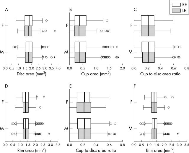
Mean values of topographic parameters for male and female participants are outlined in table 2 . No statistically significant sex dependent difference was found for any of the analysed parameters (Wilcoxon test). The analysed data show slightly larger mean RNFLT and rim volume in the right eyes of the female participants. Figure 1A–F shows box plots of the HRT measurements for men and women as well as for RE and LE including the 50% area and median in the box. Extremely high or low measurements are also shown when the data differ more than 2.5 times the SD from the median.
Table 2.
Analysis of sex related differences of least squares (LS) mean HRT optic disc measurements in normal eyes, standard deviation (SD) of LS means, differences of LS means (male):LS means (female) with non-adjusted 95% confidence interval (CI) and with significance (covariance analysis for age and sex influence on HRT measurements, Bonferoni adjusted significance level p<0.004)
| Male (n = 728), mean age 45.4 (SD 7.7) years | Female (n = 154), mean age 53.6 (SD 9.3) years | Difference (95% CI) | p Value | |
| Mean LS (SD) | Mean LS (SD) | |||
| Disc area (mm2) | ||||
| RE | 1.82 (0.40) | 1.84 (0.44) | −0.024 (−0.098 to 0.049) | 0.51 |
| LE | 1.81 (0.40) | 1.79 (0.42) | +0.023 (−0.051 to 0.098) | 0.54 |
| Cup area (mm2) | ||||
| RE | 0.44 (0.32) | 0.44 (0.34) | −0.006 (−0.065 to 0.054) | 0.85 |
| LE | 0.44 (0.33) | 0.43 (0.34) | +0.012 (−0.048 to 0.072) | 0.69 |
| Cup:disc area ratio | ||||
| RE | 0.22 (0.14) | 0.22 (0.14) | −0.001 (−0.026;0.024) | 0.95 |
| LE | 0.22 (0.13) | 0.22 (0.14) | +0.004 (−0.021 to 0.029) | 0.75 |
| Rim area (mm2) | ||||
| RE | 1.38 (0.28) | 1.40 (0.29) | −0.019 (−0.070;0.032) | 0.47 |
| LE | 1.37 (0.29) | 1.36 (0.28) | +0.011 (−0.038 to 0.061) | 0.65 |
| Mean RNFLT (mm)* | ||||
| RE | 0.261 (0.067) | 0.277 (0.070) | −0.016 (−0.028 to −0.004) | 0.011 |
| LE | 0.251 (0.066) | 0.257 (0.069) | −0.006 (−0.018 to 0.006) | 0.34 |
| Rim volume (mm3) | ||||
| RE | 0.37 (0.14) | 0.40 (0.14) | −0.032 (−0.058 to −0.008) | 0.001† |
| LE | 0.36 (0.13) | 0.37 (0.13) | −0.012 (−0.035 to 0.012) | 0.33 |
*Mean retinal nerve fibre layer thickness (HRT).
†Significant.
Figure 1.
Box plots of topographic measurements by HRT I in 1764 normal eyes with 50% area box, median in the box, minimum and maximum. Mean RNFLT (E) and rim volume (F) showed significant interocular difference (p<0.001).
To access possible age dependent changes in this cross sectional setting, the mean topographic parameters of the age quartiles were compared, as a regression analysis was not suitable for the observed data because of low correlation coefficients (R2). The analysed data were separated by age quartiles. The age range was 35–40 years in the first quartile (n = 222), 40–45 years in the second quartile (n = 221), 45–52 years in the third quartile (n = 218), and 52–70 years in the last quartile (n = 221). As shown in table 3 , no significant changes were found for cup area and cup to disc area ratio between the quartiles for the means of the analysed topographic parameters. A decrease in mean RNFLT with age was noticed in right eyes only (p = 0.04) but did not reach significance with a Bonferoni adjusted significance level of 0.05/12 = 0.0042.
Table 3.
Mean (SD) HRT optic disc parameters and difference between the age quartiles in optic nerve head measurements by HRT I in normal eyes. Difference between the first and last quartile with statistical significance (p) (Mann-Whitney U test), r2 for linear regression analysis. The age range of the quartiles was 35−40 years in the first quartile, 40−45 years in the second quartile, 45−52 years in the third quartile, 52−70 years in the last quartile
| 882 participants, mean age 46.8 (SD 8.6) years; 1764 normal eyes | |||||||
| Age quartile 1 (208 male, 14 female) (37.5 (SD 1.7) years), mean (SD) | Age quartile 2 (198 male, 23 female) (42.6 (SD 1.5) years), mean (SD) | Age quartile 3 (191 male, 27 female) (48.3 (SD 1.9) years), mean (SD) | Age quartile 4 (131 male, 90 female) (59.1 (SD 5.0) years), mean (SD) | Difference between quartiles 1 and 4 | |||
| p value | r2 | ||||||
| Disc area (mm2) | |||||||
| RE | 1.876 (0.41) | 1.841 (0.36) | 1.818 (0.39) | 1.771 (0.42) | +0.105 | 0.011 | 0.013 |
| LE | 1.808 (0.41) | 1.818 (0.36) | 1.833 (0.45) | 1.779 (0.37) | +0.029 | 0.755 | 0.002 |
| Cup area (mm2) | |||||||
| RE | 0.467 (0.32) | 0.439 (0.31) | 0.418 (0.30) | 0.435 (0.34) | +0.031 | 0.178 | 0.002 |
| LE | 0.429 (0.32) | 0.442 (0.31) | 0.441 (0.34) | 0.431 (0.32) | −0.029 | 0.953 | 0.001 |
| Cup:disc area ratio | |||||||
| RE | 0.233 (0.13) | 0.224 (0.13) | 0.216 (0.13) | 0.226 (0.15) | +0.007 | 0.436 | <0.001 |
| LE | 0.221 (0.13) | 0.228 (0.13) | 0.223 (0.14) | 0.227 (0.14) | −0.006 | 0.627 | <0.001 |
| Rim area (mm2) | |||||||
| RE | 1.409 (0.29) | 1.402 (0.24) | 1.400 (0.27) | 1.336 (0.29) | +0.073 | 0.015 | 0.013 |
| LE | 1.378 (0.26) | 1.376 (0.24) | 1.392 (0.30) | 1.348 (0.26) | +0.031 | 0.400 | 0.002 |
| Mean RNFLT (mm)* | |||||||
| RE | 0.269 (0.066) | 0.269 (0.064) | 0.264 (0.068) | 0.251 (0.067) | +0.019 | 0.004† | 0.011 |
| LE | 0.256 (0.063) | 0.255 (0.065) | 0.249 (0.068) | 0.247 (0.064) | +0.009 | 0.162 | 0.004 |
| Rim volume (mm3) | |||||||
| RE | 0.391 (0.14) | 0.389 (0.14) | 0.384 (0.16) | 0.356 (0.12) | +0.035 | 0.011 | 0.012 |
| LE | 0.368 (0.13) | 0.362 (0.12) | 0.362 (0.13) | 0.351 (0.12) | +0.017 | 0.197 | 0.002 |
*Mean retinal nerve fibre layer thickness (HRT).
†Significant.
DISCUSSION
Optical nerve head topographies measured by HRT I in 1764 normal eyes of 882 healthy participants were investigated. To date the cumulative normalised rim to disc ratio curve is in use for comparative classification of HRTI (version 2.01) measurements. This normalised data curve relies on ONH topographies of 100 eyes of 100 adults with a mean age of 36 (SD 12) years (range 9–67 years). 4 As the presented data in this paper rely on a much larger number of individuals with a higher mean age of 46.8 years (more realistic for a glaucoma patient) 14 the measurements may permit an improved automatic classification of HRT measurements in a future screening setting.
Measurements with the more recent HRT II are comparable with measurements with HRT I, except for normalised parameters, which are software depending. The clinical ONH classification included in HRT II software is based on data from 80 normal subjects and 51 patients with early glaucoma. 15
Statistically significant intraindividual differences for mean RNFLT and rim volume with lower values in the left eyes (table 1 ) were found. The observed differences in both parameters are about one sixth of the standard deviation and are not of clinical importance. In contrast, Ghergel et al found lower values for RNFLT in right normal eyes (n = 157, mean age 47.8 years) with HRT. 7 We observed significant sex related differences for mean rim volume of right eyes with higher measurements in women. The difference observed is about one sixth of the standard deviation measured. For left eyes a similar difference was not statistically significant. Ramrattan et al found significantly lower values for disc area and rim area in women (mean age 69 years) using the stereoscopic image analyser. 10 We were not able to confirm their observations. Gundersen et al found larger cup area values in women with HRT (n = 225). His findings were not statistically significant. 8 The discussed intraindividual and sex related differences are very small and do not seem to affect the evaluation of optic nerve heads in the clinical practice.
In this study we discovered statistically significant age related differences in right eyes. As shown in table 3 , the average age of women was higher than the majority of male participants. Since no significant sex related differences were observed, we assume that the age related analysis is not markedly influenced by the higher percentage of women in the fourth age quartile. Left eyes only showed a tendency to a smaller disc area, rim area, mean RNFLT, and rim volume in older volunteers. The reason for this remains unclear and may be elucidated by further studies with even larger numbers of individuals. An age related retinal nerve fibre loss in normal eyes was described by other authors. 16– 18 This may be of clinical relevance for the interpretation of optic nerve head topographies in elderly individuals. If decreasing values for rim area, rim volume, and mean RNFLT in this study were a sign for retinal nerve fibre loss with age, the cup area should consequently have increased with age. This, however, was not the case in our study population. Our data suggest stability of the cup area with age in healthy eyes. Longitudinal studies of normal eyes will help to evaluate changes in optic nerve head topography with age.
The definition for micro- and macropapillae describes abnormality rather than ONH pathology. This is clinically relevant for the evaluation of the cup volume in hyperopic and myopic eyes with suspected glaucoma, as the cup volume is related to the disc area. Adults with micropapillae should have a smaller excavation, and those with macropapillae may have a larger excavation even without glaucoma.
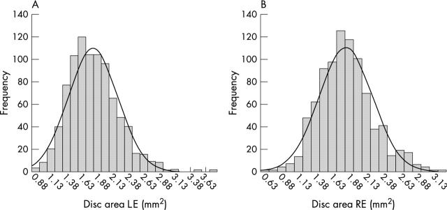
According to our findings from this large population of 1764 eyes we propose definitions for micro- and macropapillae from HRT measurements (fig 2 ). Optic discs with HRT determined disc area values below the 2.5 percentile (1.14 mm2) may be defined as micropapillae. Optic discs with disc area values above the 97.5 percentile (2.71 mm2) may be defined as macropapillae.
Figure 2.
Measurement of optic disc RE and LE area by HRT I in 1764 normal eyes. 2.5th percentile 1.14 mm2 (micropapillae); 97.5th percentile 2.71 mm2 (macropapillae).
Earlier studies using HRT in normal eyes (see table 4 ) found larger mean values for topographic optic disc parameters. The findings for disc area vary between 1.87 mm2 and 2.47 mm2. Consequently other optic disc parameters are also subject to variation. The large variation may reflect morphological differences of the populations studied but could be due to systematic measurement errors. Keratometry readings can affect the magnification error of the HRT 19 and thus influence the topographic measurements. The various ethnicities of the studied individuals and the study recruitment seem to be of minor importance. Concerning the mean topographic values our results are similar to the findings of Ghergel 7 and Gundersen 8 and in contrast to the findings of Nakamura 20 and Iester, 9 who found larger disc cups and rim areas. However, both studies 9, 20 did not use the 2.01 HRT software. Calculations with older HRT software may cause systematic differences in cup and rim values but should not alter disc area values. 6 Differences in disc size most likely reflect observer dependent drawing of the contour line defining the optic disc margin. 21 Further investigation is needed to explain the divergent results of basic topographic optic disc parameters in normal eyes.
Earlier studies using stereo photographic measurements or other techniques in normal eyes (see table 4 ) found larger mean values for topographic optic disc parameters. The findings for disc area vary between 1.87 mm2 and 3.09 mm2. 4, 7– 10, 20, 22, 23 The known mean magnification error of the HRT is less than 5% 19, 24– 26 and does not explain the discordant results.
Our data are a new basis for comparison of optic disc characteristics in healthy eyes with glaucomatous eyes. Whether these data are representative for an even larger population remains to be seen.
Abbreviations
HRT, Heidelberg retina tomograph
ONH, optic nerve head
RNFLT, retinal nerve fibre layer thickness
REFERENCES
- 1. Airaksinen PJ, Drance SM. Neuroretinal rim area and retinal nerve fibre layer in glaucoma. Arch Ophthal 1985;103:203–4. [DOI] [PubMed] [Google Scholar]
- 2. Jonas JB, Budde WM, Panda-Jonas S. Ophthalmoscopic evaluation of the optic nerve head. Surv Ophthalmol 1999;43:293–320. [DOI] [PubMed] [Google Scholar]
- 3. Anton A, Yamagishi N, Zangwill L, et al. Mapping structural to functional damage in glaucoma with standard automated perimetry and confocal scanning laser ophthalmoscopy. Am J Ophthalmol 1998;125:436–46. [DOI] [PubMed] [Google Scholar]
- 4. Bartz-Schmidt KU, Sengersdorf A, Esser P, et al. The cumulative normalised rim/disc area ratio curve. Graefes Arch Clin Exp Ophthalmol 1996;234:227–31. [DOI] [PubMed] [Google Scholar]
- 5. Caprioli J. Discrimination between normal and glaucomatous eyes. Invest Ophthalmol Vis Sci 1992;33:153–9. [PubMed] [Google Scholar]
- 6. Jonescu-Cuypers CP, Thumann G, Hilgers RD, et al. Long-term fluctuations of the normalised rim/disc area ratio quotient in normal eyes. Graefes Arch Clin Exp Ophthalmol 1999;237:181–6. [DOI] [PubMed] [Google Scholar]
- 7. Ghergel D, Orgül S, Prünte C, et al. Interocular differences in optic disc topographic parameters in normal subjects. Curr Eye Res 2000;20:276–82. [PubMed] [Google Scholar]
- 8. Gundersen KG, Heijl A, Bengtsson BO. Age, gender, IOP, refraction and optic disc topography in normal eyes. A cross-sectional study using raster and scanning laser tomography. Acta Ophthalmol Scand 1998;76:170–5. [DOI] [PubMed] [Google Scholar]
- 9. Iester M, Broadway DC, Mikelberg FS, et al. A comparison of healthy, ocular hypertensive, and glaucomatous optic disc topographic parameters. J Glaucoma 1997;6:363–70. [PubMed] [Google Scholar]
- 10. Ramrattan RS, Wolfs RCW, Jonas JB, et al. Determinants of optic disc characteristics in a general population. The Rotterdam Study. Ophthalmology 1999;106:1588–96. [DOI] [PubMed] [Google Scholar]
- 11. Burk ROW, Airkasinen PJ, Tuulonen A, et al. Reference plane for three-dimensional topographic optic disc analysis with the Heidelberg Retina Tomograph. ARVO Abstracts. Invest Ophthalmol Vis Sci 1995;36 (Suppl 4):627. [Google Scholar]
- 12. European Glaucoma Society. Terminologie und Handlungsrichtlinien zum Glaukom [in German]. Italy: Dogma 1999.
- 13. Hales GD. Retooling for the 90 s or being born again in SPSS. Comput Nurs 1990;8:230–31. [PubMed] [Google Scholar]
- 14. Dielemans I, Vingerling JR, Wolfs RC, et al. The prevalence of primary open-angle glaucoma in a population-based study in The Netherlands. The Rotterdam Study. Ophthalmology 1994;101:1851–5. [DOI] [PubMed] [Google Scholar]
- 15. Wollstein G, Garway-Heath DF, Hitchings RA. Identification of early glaucoma cases with the scanning laser ophthalmoscope. Ophthalmology 1998;105:1557–63. [DOI] [PubMed] [Google Scholar]
- 16. Balaszi AG, Rootman J, Drance SM, et al. The effect of age on the nerve fibre population of the human optic nerve. Am J Ophthalmol 1984;97:760–6. [DOI] [PubMed] [Google Scholar]
- 17. Jonas JB, Nguyen NX, Naumann GOH. The retinal nerve fiber layer in normal eyes. Ophthalmology 1989;96:627–32. [DOI] [PubMed] [Google Scholar]
- 18. Jonas JB, Schmidt AM, Muller Bergh JA, et al. Human optic nerve fiber count and optic disc size. Invest Ophthalmol Vis Sci 1992;33:2012–18. [PubMed] [Google Scholar]
- 19. Garway-Heath DF, Rudnicka AR, Lowe T, et al. Measurement of optic disc size: equivalence of methods to correct for ocular magnification. Br J Ophthalmol 1998;82:643–9. [DOI] [PMC free article] [PubMed] [Google Scholar]
- 20. Nakamura H, Maeda T, Suzuki Y, et al. Scanning laser tomography to evaluate optic discs of normal eyes. Jpn J Ophthal 1999;43:410–14. [DOI] [PubMed] [Google Scholar]
- 21. Diestelhorst M, Burk ROW, Garway-Heath DF, et al. Observer dependent diagnostic variability of optic nerve head measurements with confocal laser scanning tomography. [Abstract] Invest Ophthalmol Vis Sci 2002;43:E1014. [Google Scholar]
- 22. Broadway DC, Drance SM, Parfitt CM, et al. The ability of scanning laser ophthalmoscopy to identify various glaucomatous optic disc appearances. Am J Ophthalmol 1998;125:593–4. [DOI] [PubMed] [Google Scholar]
- 23. Budde WM, Jonas JB, Martus P, et al. Influence of optic disc size on neuroretinal rim shape in healthy eyes. J Glaucoma 2000;9:357–62. [DOI] [PubMed] [Google Scholar]
- 24. Bartz-Schmidt KU, Weber J, Heimann K. Validity of two-dimensional data obtained with the Heidelberg Retina Tomograph as verified by direct measurements in normal optic nerve heads. Ger J Ophthalmol 1994;3:400–5. [PubMed] [Google Scholar]
- 25. Janknecht P, Funk J. Optic nerve head analyzer and Heidelberg retina tomograph: relative error and reproducibility of topographic measurements in a model eye with simulated cataract. Graefes Arch Clin Exp Ophthalmol 1995;233:523–9. [DOI] [PubMed] [Google Scholar]
- 26. Zangwill L, Irak I, Berry CC, et al. Effect of cataract and pupil size on image quality with confocal scanning laser ophthalmoscopy. Arch Ophthalmol 1997;115:983–90. [DOI] [PubMed] [Google Scholar]


