Strabismus is a common association in patients with craniosynostosis or craniofacial dysostosis (60–70%).1–3 V pattern exotropia is the most common ocular motility problem.
Various theories have been proposed to explain the cause of the V pattern and surgical attempts to correct it with weakening procedures of the inferior oblique have been disappointing.2,3
This is a case report of one child with this disorder who underwent orbital computed tomography (CT) scans and had a marked improvement of the V pattern following strabismus surgery based on the CT findings.
CASE REPORT
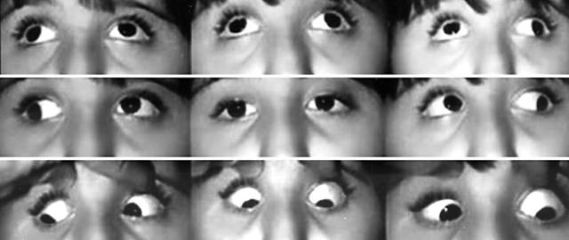
This child with craniosynostosis had undergone six previous cranial surgeries. She had three strabismus surgical procedures including anterior transpositions of the inferior obliques in an attempt to correct a large V pattern. She presented to us with a chin up position, V pattern exotropia (60 prism dioptres), over-elevation in adduction, limitation of depression in adduction, and incomitant hypertropias in side gazes (fig 1). Objective fundus excyclotorsion was noted.
Figure 1.
Preoperative V pattern exotropia, over-elevation in adduction, under-depression in adduction in both eyes.
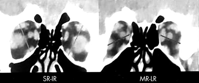
Orbital imaging demonstrated that all extraocular muscles in each eye were present, normal in size and shape but anatomically displaced. The extraocular muscles in the left eye were rotated clockwise and in the right eye were rotated counterclockwise (fig 2). Ineffectiveness of inferior oblique weakening procedures and the presence of muscle heterotopy led us to consider that the over-elevation in adduction was most likely related to the anatomical displacement of the rectus muscles.
Figure 2.
Preoperative coronal views on CT scan of both orbits showing evidence of rectus muscle heterotopy. A vertical line joining the centre of the belly of the vertical rectus muscles shows the relative temporal displacement of the superior rectus muscle compared to the nasal displacement of the inferior rectus muscle. A horizontal line joining the centre of the belly of the horizontal rectus muscles shows the relative inferior displacement of the lateral rectus muscle compared to superior displacement of the medial rectus muscle.
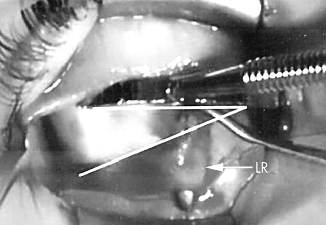
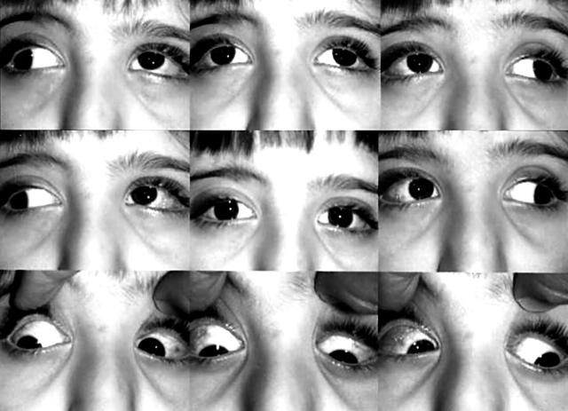
Surgical exploration confirmed muscle heterotopy. The lateral recti were found slanting inferiorly (fig 3). Repositioning of the lateral recti superiorly to a more horizontal position and suturing the superior border of the muscle belly to the adjacent sclera about 18 mm from the limbus using a non-absorbable suture was the first surgical procedure performed by us on this patient. This led to some improvement of the V pattern. This was followed by recession and nasal repositioning of the superior recti suturing the nasal border of the muscle belly to the adjacent sclera about 18 mm from the limbus using a non-absorbable suture. This achieved good alignment in the primary position and eliminated the anomalous chin up position, markedly reduced the V pattern, eliminated the over-elevation in adduction, and improved depression in adduction (fig 4).
Figure 3.
Intraoperative photograph. The left eye is adducted with a muscle hook placed under the lateral rectus muscle (LR). The lower line highlights the downward slanting of the left lateral rectus muscle. A curved ruler is used to show the normal horizontal path of the lateral rectus muscle.
Figure 4.
Postoperative clinical pictures showing improvement in V pattern exotropia with improvement in versions in adduction.
COMMENT
V pattern strabismus in craniosynostosis may be related to anatomical malposition of the rectus muscles. This may be documented by orbital imaging, which could also aid in planning the surgical approach. In these cases the overelevation in adduction and under depression in adduction may be due to the anatomical displacement of the rectus muscles.2
References
- 1.Coats DK, Paysse EA, Stager DR. Surgical management of V-pattern strabismus and oblique dysfunction in craniofacial dysostosis. JAAPOS 2000;4:338–42. [DOI] [PubMed] [Google Scholar]
- 2.Clark RA, Miller JM, Rosenbaum AL, et al. Heterotopic muscle pulleys or oblique muscle dysfunction? JAAPOS 1998;2:17–25. [DOI] [PubMed] [Google Scholar]
- 3.Limon de Brown , Monasterio FO, Feldman MS. Strabismus in plagiocephaly. J Pediatr Ophthalmol Strabismus 1998;25:180–90. [DOI] [PubMed] [Google Scholar]


