Abstract
Aim: To evaluate the complications of periocular full thickness skin grafts (FTSG) in patients treated with Mohs’ micrographic surgery (MMS) for periocular malignancy.
Method: This prospective, multicentre case series included all patients in Australia treated with MMS for periocular malignancy followed by reconstruction with FTSG, who were monitored by the Skin and Cancer Foundation, between 1993 and 1999. The parameters recorded were patient demographics, reason for referral, histological classification of malignancy and evidence of perineural invasion, duration of tumour, site, recurrences prior to MMS, preoperative tumour size, and postoperative defect size. FTSG donor sites included upper lid, preauricular, retroauricular, inner brachial, and supraclavicular. The primary outcome measures were FTSG host site complications (partial/complete graft failure, graft infection, acute bleeding/haematoma, graft hypertrophy, and graft contracture).
Results: 397 patients (229 males, 168 females), mean age 60 (SD 15) years (range 20–91 years). 92.7% were diagnosed with basal call carcinoma, 2.0% with Bowen’s disease, and 3.3% with squamous cell carcinoma. Medial canthus was involved in 66.5% of patients, lower eyelid in 28.0%, and upper eyelid in 5.5%. Postoperative complications were recorded in 62 patients (15.6% of all patients), and consisted of graft hypertrophy (45.1% of complications), graft contraction (29.1%), and partial graft failure (12.9%). The only statistically significant association found was a higher rate of graft hypertrophy in medial canthal reconstruction (p = 0.007).
Conclusion: Host site complications of periocular FTSG are not common. Graft hypertrophy accounted for most complications and was more common in the medial canthal area. No other variables such as patient demographics, tumour characteristics, or donor site factors were associated with a higher risk of complications.
Keywords: complications, contracture, full thickness skin grafts, hypertrophy
Although known for almost 3000 years, full thickness skin grafts (FTSG) were introduced to the Western world only in the nineteenth century.1,2 FTSG are now frequently used by plastic surgeons as anterior lamella substitutes in eyelid reconstruction surgery, providing tissue with similar colour, texture, and thickness.1,2 The periocular area is considered a suitable host, having a rich vascular supply for capillary regrowth, as well as collagen producing fibroblasts which help in graft adherence.1,2 This successful combination of graft and host has subsequently made it widely used in eyelid reconstruction surgery after trauma, burns, and tumour removal.
This prospective study aimed to assess the incidence and characteristics of FTSG complications in a large series of patients undergoing reconstructive eyelid surgery after Mohs micrographic surgery (MMS) for periocular tumours.
METHOD
We conducted a prospective, non-comparative, multicentre, interventional case series of patients with periocular malignancy treated with MMS in Australia and monitored by the Skin and Cancer Foundation, between 1993 and 1999. All patients were treated by an accredited Mohs surgeon, using standard fresh frozen MMS techniques. Data were collected by the Skin and Cancer Foundation.
The criteria for selection were all cases with periocular malignancy treated with MMS followed by reconstruction using FTSG. Periocular was defined as medial canthus, upper eyelid, or lower eyelid. Tumours outside this region were excluded from this study.
The following parameters were recorded: patient identification number, age, sex, reason for referral, histological classification of malignancy and evidence of perineural invasion (PNI), duration of tumour, site, recurrences prior to MMS, preoperative tumour size, and postoperative defect size. Tumour and postoperative defect size were defined into eight groups based on the maximum diameter using a straight rule: 0–0.9 cm, 1–1.9 cm, 2–2.9 cm, 3–3.9 cm, 4–4.9 cm, 5–5.9 cm, 6–7.9 cm, and 8–10 cm. FTSG donor sites included upper lid (ipsilateral or contralateral), preauricular, retroauricular, inner brachial, and supraclavicular).
The primary outcome measures were FTSG host site complications up to a follow up period of 6 months. All cases were assessed for skin graft complications including: partial/complete graft failure, graft infection, acute bleeding/haematoma, graft hypertrophy (defined as more than 2 mm elevation of the graft from surrounding skin), and graft contracture (defined as greater than 50% contraction of the FTSG), and “trapdoor” contracture (graft elevated centrally and depressed at the circumference).
Statistical analysis
Univariate log binomial regression analysis was performed on each of the potential binary variable complications. The final multivariate model included the variables with a p value of less than 0.2 in the univariate analysis. Significance for the final model was assessed at the 5% level. The results were reported using relative risks and 95% confidence intervals. Fisher’s exact test was used when individual types of complications were evaluated, and significance was assessed at the 5% level. All analyses were performed using SAS version 8.2 (SAS Institute Inc, Cary, NC, USA).
RESULTS
The study included 397 patients who underwent MMS followed by reconstruction using FTSG, between 1993 and 1999. There were 229 males (57.7%) and 168 females (42.3%), mean (standard deviation) age 60 (SD 15) years (range 20–91 years).
A total of 368 patients (92.7%) were diagnosed with basal call carcinoma (BCC), eight patients (2.0%) with Bowen’s disease, and 13 patients (3.3%) with squamous cell carcinoma. In eight patients (2.0%) the details of tumour type were not available.
The tumour involved the lower eyelid in 111 patients (28.0%), the upper eyelid in 22 patients (5.5%), and medial canthus in 264 patients (66.5%). The majority of tumours (88.6%) were smaller than 2 cm and most of the post-surgical defects (81.6%) were smaller than 3 cm (table 1).
Table 1.
Periocular tumours size and postoperative defect size
| Tumour/defect size (cm) | Number of patients with tumour (%) | Number of patients with defect (%) |
| <1 | 147 (37.0) | 8 (0.2) |
| 1–1.9 | 205 (51.6) | 183 (46.1) |
| 2–2.9 | 29 (7.3) | 133 (33.5) |
| 3–3.9 | 8 (2.0) | 49 (12.3) |
| 4–4.9 | 2 (0.5) | 15 (3.8) |
| 5–5.9 | 0 | 4 (1.0) |
| >6 | 0 | 2 (0.5) |
| No details available | 6 (1.5) | 3 (0.8) |
The most common site for FTSG harvesting was the supraclavicular area (44.6%), followed by upper lid (20.9%), retroauricular (16.6%), preauricular (2.5%), and inner brachial (1.3%). In 56 patients (14.1%), no details were available regarding FTSG site harvesting.
FTSG complications
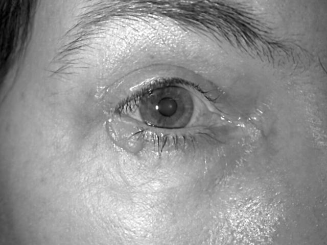
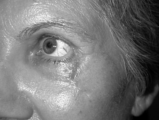
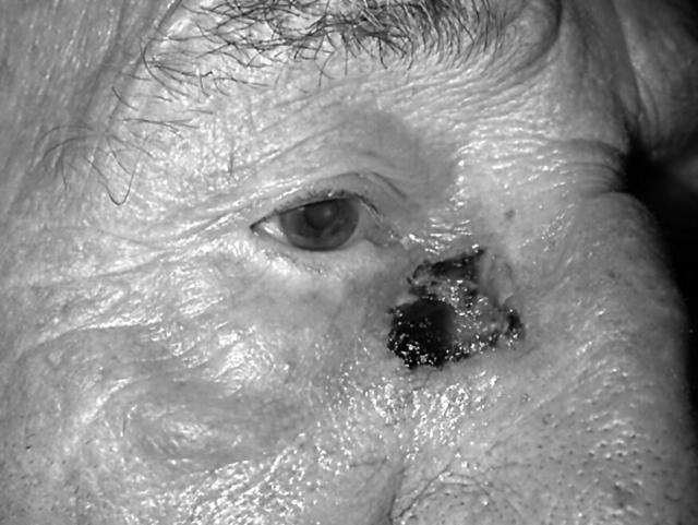
Postoperative complications were recorded in 62 patients (15.6%) (table 2). The most common complications were graft hypertrophy (fig 1) (45.1% of all complications), graft contraction (fig 2) (29.1%), and partial graft failure (fig 3) (12.9%).
Table 2.
Postoperative complications after MMS with FTSG according to periocular tumour site
| Type of graft complication | Medial tumours (%)* n = 264 | Lower lid tumours (%)* n = 111 | Upper lid tumours (%)* n = 22 | Total number (%)* n = 397 |
| Hypertrophy | 26 (41.9)† | 2 (3.2) | 28 (45.1) | |
| Contracture | 2 (3.2) | 2 (3.2) | 4 (6.5) | |
| Contracture+ectropion | 2 (3.2) | 2 (3.2) | 1 (1.6) | 5 (8.1) |
| Trapdoor contraction | 6 (9.6) | 1 (1.6) | 7 (11.3) | |
| Web contracture | 2 (3.2) | 2 (3.2) | ||
| Partial failure | 4 (6.4) | 4 (6.4) | 8 (12.9) | |
| Complete failure | 1 (1.6) | 1 (1.6) | ||
| Infection | 3 (4.9) | 1 (1.6) | 4 (6.5) | |
| Hematoma | 2 (3.2) | 1 (1.6) | 3 (4.8) | |
| Total number (%) | 48 (77.4) | 13 (21) | 1 (1.6) | 62 (100) |
*% from total number of complications.
†p = 0.007.
Figure 1.
Graft hypertrophy after right lower lid reconstruction. Reproduced with the patient’s permission.
Figure 2.
Graft contracture after left lateral lower lid reconstruction. Reproduced with the patient’s permission.
Figure 3.
Partial graft failure after medial canthal defect reconstruction. Reproduced with the patient’s permission.
Statistical analysis was performed to identify association between the variables (age, sex, reason for referral, histological classification of malignancy and evidence of PNI, duration of tumour, tumour site, recurrences prior to MMS, surgeon, preoperative tumour size, and postoperative defect size, FTSG donor sites) and postoperative complications (partial or complete graft failure, graft infection, acute bleeding/haematoma, graft hypertrophy, and graft contracture). No association was found between the complications (all complications together as a group and each of them separately) and any of the following variables: age, sex, reason for referral, histological subtype, PNI, duration of tumour, prior recurrence, surgeon, preoperative tumour size, postoperative defect, or the harvesting site for the FTSG. The only statistically significant association found was between medial canthal tumour location and the postoperative graft hypertrophy (p = 0.007). No associations were found between any of the other complications and tumour location.
The treatment modalities used for postoperative FTSG complications included intralesional steroid injection in 34 patients (12 patients with graft contracture, 21 with graft hypertrophy, and one with partial graft failure), topical steroids (in one patient with graft hypertrophy), and CO2 laser treatment combined with intralesional steroid injection (in one patient with graft hypertrophy). Hence, 36 (9%) patients out of 397 who underwent FTSG required treatment for graft complications. Data regarding the long term effect of these treatments or donor site complications were not available.
DISCUSSION
Full thickness skin grafts are composed of epidermis and the entire dermis. When used in periocular reconstruction they are usually harvested from several possible donor sites (upper lid, preauricular, retroauricular, neck, clavicular and supraclavicular, and inner brachial area) yielding different graft thicknesses accordingly.1–3 The main advantages of FTSG are availability, low metabolic requirement and resistance to trauma.3 Skin grafts go through a unique process of healing in the host site.1,2 The first phase, which lasts 24 hours, is an ischaemic stage (called “plasmatic imbibition”), followed by an oedematous stage in which the graft gains up to 40% in weight, and finally revascularisation of the graft (“inosculation”), which becomes apparent within 48–72 hours after grafting. The blood supply to the graft comes from recipient bed (“bridging phenomenon”).1,2 Alteration in any of these stages may result in graft failure and complications.
Postoperative host site complications of full thickness skin grafting may arise shortly after the operation and result in partial or complete graft failure, or develop gradually and cause functional and cosmetic problems later.1–3 These complications have not been specifically studied in the periocular area. Our series, to the best of our knowledge, is the largest reported prospective series of periocular FTSG complications.
The early complications are mainly bleeding with haematoma formation beneath the graft, infection, or seroma formation. These complications may prevent graft adherence to the underlying wound bed, prolong the ischaemic phase, compromise the graft’s vascular supply, and result in graft failure. Additionally, graft movement or shear forces may also lead to graft failure by disrupting the attachment of the graft to the wound bed.1,2 The rich facial vascular supply makes postoperative infection uncommon, but on the other hand, increases the risk of haematoma formation, especially in patients treated with non-steroidal anti-inflammatory drugs and anticoagulants. These patients are at risk for short term complications and should be monitored accordingly.
The long term complications are mainly cosmetic or functional and result from colour and texture mismatch, hyper- or hypopigmentation, graft hypertrophy, and graft contraction.1–3 A successful skin graft is usually light pink in colour in the early postoperative period, but it can sometimes be red, dark blue, or purple.1,2 A black graft signifies partial or total graft failure and necrosis. Colour mismatch and pigmentation differences are generally temporary and improve gradually, but may, on occasion, be permanent and require intervention with dermabrasion or laser resurfacing.1,2 Colour mismatch complications were not reported in our study, and it is possible they were underreported.
Graft contraction is secondary to centripetal movement of the unapposed elastic fibres, resulting in variable degrees of shrinkage. The factors influencing shrinkage are mainly elasticity of the donor site and graft thickness.1,2 Graft contraction is believed to be more prominent as the thickness of the graft decreases,3–5 but it is generally thought that FTSG contract minimally in humans.6 Stephenson et al7 on the other hand, have recently published a study evaluating patterns of FTSG contractions in humans. They found that in the presence of infection, the graft contracted to almost half the initial size, and in cases where there was no infection, the graft contracted by one third. In addition, more contraction occurred in grafts placed in the nasal and periorbital areas compared with the temple and scalp; however, they found no significant difference in contraction between donor sites. In our study, there were only 18 cases of significant graft contracture (4.5% of all patients and 29% of all complications, table 2). Although the majority of the contracture cases occurred in medial canthal grafts (12 cases out of 18), this was not statistically significant, and was explained by the relatively higher number of medial tumours treated (264 cases out of 397; 66.5%). In accordance with Stephenson’s study,7 we found no significant difference in graft contraction between the different donor sites for FTSG harvesting. Although our series did not find a significant rate of lid malposition, it should be noted that contracture of FTSG in the periocular region carries the potential for added morbidity in the form of ectropion or retraction.
Graft hypertrophy was defined in our study as more than a 2 mm elevation of the graft from the surrounding skin. It occurred in 28 patients (7.0% of all cases and 45.1% of all complications, table 2). Graft hypertrophy was most common in medial canthal defect reconstruction compared with other periocular sites, and this difference was statistically significant (p = 0.007). No other tumour related or donor site variables in our study were associated with a higher percentage of graft hypertrophy. The exact mechanism for graft hypertrophy is not fully understood, and it probably represents aberration in the process of wound healing, which includes cell proliferation, inflammation, and increased synthesis of cytokines and extracellular matrix proteins. This may be a similar process to that of hypertrophic scars and keloids formation.8 The larger number of hypertrophic grafts in the medial canthal area may be due to a higher degree of cellular proliferation and wound healing in this area. In addition, the medial canthal area is a confluence of three aesthetic units (medial canthus, upper lid, and lower lid) with different degrees of skin thickness, texture, colour, and contour. It is believed that replacing lost skin with grafts of similar histology, texture, and thickness may have favourable cosmetic results.11–13 Periocular tumours in this concave area usually extend over several units, and so reconstruction with a single skin graft may not respect the boundaries of these units. The resulting scar in this area may be hypertrophied or cause contracture.
Although there are no studies which evaluated the role of FTSG size relative to the defect and the development of complications such as contracture and hypertrophy, it is possible that oversizing the graft is likely to reduce the risk of these complications. In our study, the graft size data were not available, and so we were not able to draw any relevant conclusions. Skin graft thickness may also play an important role in postoperative graft contraction, which appears to occur more frequently as the thickness decreases.3–5 Our study is based on results of a number of different clinicians who were all experienced reconstructive surgeons and it is probable that the grafts were oversized to some degree and were not overthinned. In addition, although there were no data to correlate between the rate of complication and graft size or thickness, we found no correlation between these complications and any individual surgeon.
There are no studies which have specifically evaluated treatments for hypertrophic skin grafts, and the current treatment modalities are mainly based on those used for treating hypertrophic scars and keloids. The conservative options include observation, pressure garments, massage, and silicone gel sheets.8 Intralesional injection of steroids, as performed in 22 of our patients, has been shown to be effective in patients with keloids and hypertrophic scars,8 but has not been specifically studied in hypertrophic skin grafts. The most common agents used are triamcinolone acetonide and triamcinolone diacetate which may be combined with pulsed dye lasers.9,10 Steroid injection may be associated with local side effects such as pain, dermal atrophy, necrosis, ulceration, and hypopigmentation.8,9 Other possible treatment modalities are dermabrasion (such as aluminium oxide crystals, acids, liquid nitrogen, and others) or laser CO2 resurfacing which may also improve texture and colour abnormalities.9 The last two methods are also mainly used for scar revision, and there are no studies of their effectiveness in skin graft hypertrophy treatment. Conservative measures such as observation in cases of graft hypertrophy may be a reasonable treatment option which may result in improvement or resolution in many cases; however, although anecdotally this is the experience of many clinicians, there are no studies to support this option. In cases where the graft is causing considerable hypertrophy, it would seem reasonable to use the treatment modalities described. Many of the cases in our series with hypertrophy and contracture were managed conservatively. It is also possible that some of those complications which received treatment would have improved or resolved completely without any intervention. Our study, however, was primarily aimed at assessing the incidence of graft complications rather than evaluating the efficacy of different treatment modalities.
In conclusion, although we have identified several possible host site complications associated with periocular FTSG, they are not common and often do not require active treatment, making FTSG a good choice in eyelid reconstructive surgery. Skin graft hypertrophy accounted for the majority of complications and was more frequent in the medial canthal region. No other variables such as patient demographics, tumour characteristics, or donor site factors were associated with a higher risk of complications. It may be prudent for the clinician to inform patients undergoing periocular FTSG that, in addition to the normal evolution in colour and thickness in the short term, there is small risk of graft hypertrophy or contracture which may require additional treatment.
Acknowledgments
The authors wish to thank the Skin and Cancer Foundation Australia and the participating Mohs surgeons for their generosity in providing the data for this research. The Mohs surgeons involved were Drs Phillip Artemi, John Coates, Brian De’Ambrosis, Timothy Elliott, Gregory Goodman, Irene Grigoris, Dudley Hill, Shyamala Huilgol, Michelle Hunt, David Leslie, Robert Paver, Shawn Richards, William Ryman, Robert Salmon, Margaret Stewart, Howard Studniberg, Carl Vinciullo, and Perry Wilson.
The authors also wish to thank Emmae Ramsay (Statistician), Department of Public Health University of Adelaide, for her help and advice in the statistical analysis of data.
Abbreviations
FTSG, full thickness skin graft
MMS, Mohs’ micrographic surgery
PNI, perineural invasion
REFERENCES
- 1.Johnson TM, Ratner D, Nelson BR. Soft tissue reconstruction with skin grafting. J Am Acad Dermatology 1992;27:151–65. [DOI] [PubMed] [Google Scholar]
- 2.Ratner D . Skin grafting. From here to there. Dermatol Clin 1998;16:75–90. [DOI] [PubMed] [Google Scholar]
- 3.Valencia IC, Falabella AF, Eaglstein WH. Skin grafting. Dermatol Clin 2000;18:521–32. [DOI] [PubMed] [Google Scholar]
- 4.Mir Y, Mir L. Biology of the skin graft. Plast Reconstr Surg 1951;8:378. [DOI] [PubMed] [Google Scholar]
- 5.Rudolph R . The effect of skin graft preparation on wound contraction. Surg Gynecol Obstet 1976;142:49–56. [PubMed] [Google Scholar]
- 6.Place MJ, Herber SC, Hardesry RA. In: Aston SJ, Beasley RW, Thorne CH, eds. Grabb and Smith’s plastic surgery. 5th edition. Philadelphia: Lippincott-Raven, 1987.
- 7.Stephenson AJ, Griffiths RW, La Hausse-Brown TP. Patterns of contraction in human full thickness skin grafts. Br J Plast Surg 2000;53:397–402. [DOI] [PubMed] [Google Scholar]
- 8.Tredget EE, Nedelec B, Scott PG, et al. Hypertrophic scars, keloids and contractures. The cellular and molecular basis for therapy. Surg Clin North Am 1997;77:701–30. [DOI] [PubMed] [Google Scholar]
- 9.Moran ML. Scar revision. Otolaryngol Clin North Am 2001;34:767–80. [DOI] [PubMed] [Google Scholar]
- 10.Alster T . Laser scar revision: comparison study of 585-nm pulsed dye laser with and without intralesional corticosteroids. Dermatol Surg 2003;29:25–9. [DOI] [PubMed] [Google Scholar]
- 11.Gonzales-Ulloa M . Restoration of the face covering by means of selected skin in regional aesthetic units. Br J Plast Surg 1956;9:212. [DOI] [PubMed] [Google Scholar]
- 12.Maillard GF, Clavel PR. Aesthetic units in skin grafting of the face. Ann Plast Surg 1991;26:347. [DOI] [PubMed] [Google Scholar]
- 13.Harris GJ, Logani SC. Multiple aesthetic unit flaps for medial canthal reconstruction. Ophthal Plast Reconstr Surg 1998;14:352–9. [DOI] [PubMed] [Google Scholar]


