A true pterygium is a degenerative and hyperplastic process in which the cornea is invaded by a triangular fold of bulbar conjunctiva. Duke-Elder states that the pterygium when single is almost invariably found on the nasal side.1 The literature on pterygium is abundant and almost from the beginning the emphasis has been placed on its location on the nasal side.
Squamous cell neoplasia of the conjunctiva is relatively uncommon and can masquerade as common, but less significant, ocular surface conditions including pterygium or chronic blepharoconjunctivitis. We present a case of intraepithelial neoplasia, initially diagnosed as inflamed pterygium.
Case report
A 77 year old man, who had worked on the railways, presented with a 3 week history of redness on the outer aspect of the left eye. No history of associated pain, discharge, or watering was elicited.
His medical history included hypertension and hypercholesterolaemia under treatment.
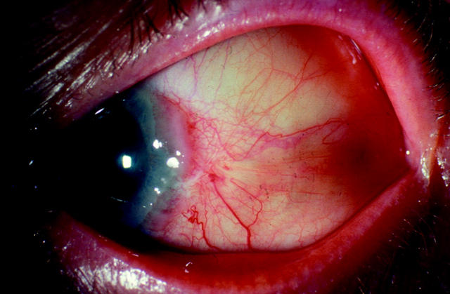
Best corrected visual acuity in each eye was 6/5. On inspection of the anterior segment, the left temporal conjunctiva showed a fleshy tissue encroaching on the temporal peripheral cornea (fig 1). The peripheral cornea showed an elevated ridge with punctate staining. The overlying conjunctiva was injected. The rest of the ocular examination was within normal limits.
Figure 1.
Left eye showing presence of a soft tissue lesion on the temporal conjunctiva encroaching on the limbus before local excision and radiotherapy.
A provisional diagnosis of inflamed pterygium of left eye was made and the patient was commenced on prednisolone 0.5% eye drops at this stage with advice to review in 2 weeks’ time.
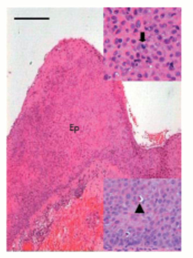
On follow up no significant change was noticed in the lesion. On further inquiry the patient gave a history of injury to left eye with hot ashes many years earlier. In view of the atypical location and the appearance of the lesion, we did an excision biopsy of the conjunctival and corneal lesion. Histopathology revealed an irregular epithelial thickening associated with dyskeratosis and full thickness dysplasia. Numerous mitotic figures, some atypical, were present throughout the epithelium (fig 2). A diagnosis of conjunctival intraepithelial neoplasia was made. Although no unequivocal evidence of invasion was seen in the multiple sections examined, fragmentation of the tissue during processing precluded confirmation of complete excision.
Figure 2.

Section through the conjunctiva stained with haematoxylin and eosin. The main figure demonstrates grossly and irregularly thickened, dysplastic epithelium (Ep; scale bar, 100 µm). The inserts show an atypical mitosis (above, arrow) and dyskeratosis (below, arrowhead) within the epithelium.
The patient was referred for further treatment to an ocular oncologist and underwent ruthenium plaque therapy followed by topical 5-fluorouracil treatment.
Comment
Temporal pterygium is reported, although Dolezalova found only one case of unilateral temporal pterygium out of 1388 Arab patients with pterygia.2 We would therefore consider this case to be atypical.
The role of pterygium in the development of ocular surface squamous neoplasia is unclear.3 Both conditions have a strong association with exposure to ultraviolet-B radiation. Sevel and Sealy’s study of 12 squamous cell carcinoma and 17 carcinoma in situ arising in 100 pterygia found that it can be difficult to distinguish a “reactive pterygium” from carcinoma in situ and malignant change should be considered in a pterygium if there is unusual evidence of invasion, extension, or if the lesion becomes particularly vascular.4
To our knowledge, the last reported case of temporal pterygium was in the 1970s.2,5 We present this case to refresh the memory and to highlight the importance of keeping an index of suspicion for squamous cell neoplasia in any atypical presentation of the more common conjunctival lesions such as pterygium.
Competing interests: none declared
References
- 1.Duke-Elder S. System of ophthalmology. Vol III, Part I. St Louis: Mosby, 1965:573–83.
- 2.Dolezalova V. Is the occurrence of a temporal pterygium really so rare? Ophthalmologica 1977;174:88–91. [DOI] [PubMed] [Google Scholar]
- 3.Lee GA, Hirst LW. Ocular surface squamous neoplasia. Surv Ophthalmol 1995;39:429–50. [DOI] [PubMed] [Google Scholar]
- 4.Sevel D, Sealy R. Pterygia and carcinoma of the conjunctiva. Trans Ophthalmol Soc UK 1968;88:567–8. [PubMed] [Google Scholar]
- 5.Awan KJ. The clinical significance of a single unilateral temporal pterygium. Can J Ophthalmol 1975;10:222–6. [PubMed] [Google Scholar]


