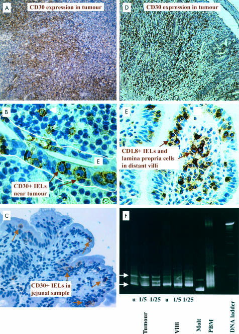
Figure 1.
Immunomorphological and genotypic observations in overt enteropathy associated T cell lymphoma (EATL) (A–C, patient No 5; D–F, patient No 6). (A) Detail of ulcerated tumour with CD30+ tumour cells in a surgical resection specimen (original magnification ×200). (B) Detail of epithelium adjacent to CD30+ tumour in (A) (original magnification ×400). Numerous intraepithelial lymphocytes express CD30 (arrows). (C) Occasional intraepithelial lymphocytes express CD30 (arrows) in a jejunal biopsy specimen (original magnification ×400). (D) Detail of ulcerated tumour with CD30+ tumour cells in a surgical resection specimen (original magnification ×100). (E) Villus a long way away from the tumour showing numerous intraepithelial and lamina propria cells (arrows) expressing CD30 (original magnification ×400). (F) Polymerase chain reaction analysis of DNA extracted from tumour and intact mucosa (villi). Samples were undiluted (u), or diluted 1/5 or 1/25; visible bands (arrows) were present in the same position of all samples, indicating that the same T cell clone was present in villi and in the tumour.

