I read with interest the report of Cruz-Correa and colleagues (Gut 2002;51:60012235088). They described three cases of collagenous colitis with mucosal tears on endoscopic insufflation and stated that as far as they were aware there were no reports in other gastrointestinal diseases. We would like to present the case of a similar mucosal tear on endoscopic insufflation in a patient with diversion colitis.
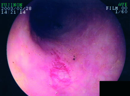
A 46 year old Japanese man presented with an acute abdomen caused by ascending colon diverticular perforation. He underwent drainage of the abdominal cavity with loop colostomy. He had been suffering from systemic lupus erythematosus and chronic renal failure for 25 years. He had received more than 90 g of oral steroid at the time of referral and was taking 10 mg/day. After operation, he was free from symptoms and gave no history of haematemesis or blood in stools. On surveillance colonoscopy, the dysfunctional colon mucosa, which was 10 cm away from the loop colostomy, was torn with slight bleeding, and the muscularis mucosal was exposed on endoscopic insufflation with air (fig 1▶). The lumen of the colon was narrowed and the remaining colon mucosa showed mild colitis with a decreased vascular pattern and oedema. The post endoscopic course was uneventful without any treatment. Routine laboratory investigations revealed: white blood cell count 10600/μl (normal range 4000–9000/μl), haemoglobin 12.2 g/dl (normal range 14–18 g/dl), haematocrit 36.1% (normal range 40–48%), blood urea nitrogen 76.4 mg/dl (normal range 9–21 mg/dl), and serum creatinine 4.29 mg/dl (normal range 0.6–1.2 mg/dl). Cultures for stool pathogens were negative.
Figure 1 .
Endoscopic insufflation of a diverted colon resulted in a mucosal tear.
Diversion colitis may occur in a part of the bowel that was previously healthy and which has been placed outside the faecal stream because of a proximal stoma.1 The mechanism of diversion colitis remains unclear but may be associated with changes in the intestinal bacterial flora, absence of essential nutrients, or intestinal toxins. In most cases, there are no symptoms, as in our case. Frisbie et al reported that mucosal erythema and friability were seen in most patients who had undergone diverting colostomy for neuropathic large bowel.2 Continuous high doses of steroids make human tissue fragile, including the colon mucosa. Taken together, these results suggest that the mucosal tear in our case may have been attributable to diversion colitis with fragile mucosa.
When performing surveillance colonoscopy for patients with a stoma, the dysfunctional colorectum must be surveyed with great care.
References
- 1.Glotzer DJ, Glick ME, Goldman H. Proctitis and colitis following diversion of the fecal stream. Gastroenterology 1981;80:438–441. [PubMed] [Google Scholar]
- 2.Frisbie JH, Ahmed N, Hirano I, et al. Diversion colitis in patients with myelopathy: clinical, endoscopic, and histopathological findings. J Spinal Cord Med 2000;23:142–9. [DOI] [PubMed] [Google Scholar]


