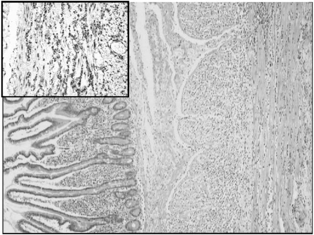
Figure 1.
Full thickness biopsy of the small intestine with haematoxylin-eosin. The section shows a normal mucosal layer of jejunum without atrophy or excessive amounts of round cells. The muscularis mucosae is also normal. In contrast, the muscularis propria shows a heavy lymphocytic infiltrate (haematoxylin-eosin). Insert: immunohistochemical stain for CD8 lymphocytes in the muscularis propria.

