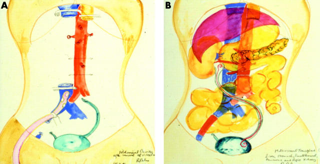
Figure 3.
Illustration of multivisceral transplantation. (A) Following preparation of the recipient for grafting. Clamps are shown on the inferior vena cava and oesophagus. All abnormal organs apart from the bladder have been excised. (B) Following grafting of the stomach, liver, pancreas, kidney, and small intestine. A stent was placed in the re-anastomosed ureter. A segment of iliac vein on the right has been replaced. Original water colours by Sir Roy Calne, with kind permission. This illustration originally appeared in Chan and colleagues.73

